8Q8H image
Deposition Date 2023-08-18
Release Date 2024-05-15
Last Version Date 2024-05-15
Entry Detail
PDB ID:
8Q8H
Keywords:
Title:
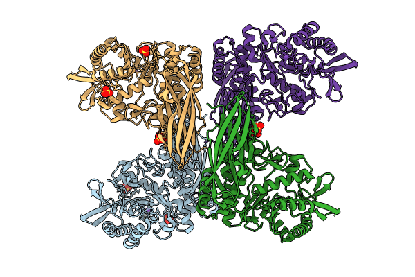
Crystal Structure of Apo beta-D-GalNAcase from Niabella aurantiaca (Structure 2)
Biological Source:
Method Details:
Experimental Method:
Resolution:
2.50 Å
R-Value Free:
0.23
R-Value Work:
0.18
R-Value Observed:
0.19
Space Group:
P 21 21 2
Macromolecular Entities
Structures with similar UniProt ID
Protein Blast
Polymer Type:polypeptide(L)
Molecule:beta-D-GalNAcase from Niabella aurantiaca DSM 17617
Chain IDs:A, B, C, D
Chain Length:536
Number of Molecules:4
Biological Source:Niabella aurantiaca DSM 17617
Primary Citation
Characterization and structural study of a novel beta-N-acetylgalactosaminidase from Niabella aurantiaca.
Febs J. 291 1439 1456 (2024)
PMID: 38129294 DOI: 10.1111/febs.17042

Abstact

We report here the identification, characterization and three-dimensional (3D) structure determination of NaNga, a newly identified β-N-acetylgalactosaminidase from the Gram-negative soil bacterium Niabella aurantiaca DSM 17617. When recombinantly expressed in Escherichia coli, the enzyme selectively cleaved 4-nitrophenyl-N-acetyl-β-d-galactosamine (pNP-β-d-GalpNAc). The X-ray crystal structure of the protein was refined to 2.5 Å and consists of an N-terminal β-sandwich domain and a (β/α)8 barrel catalytic domain. Despite a mere 22% sequence identity, the 3D structure of NaNga is similar to those previously determined for family GH123 members, suggesting it also employs the same substrate-assisted catalytic mechanism. Inhibition by N-acetyl-galactosamine thiazoline (GalNAc-thiazoline) supports the suggested mechanism. A phylogenetic analysis of its proximal sequence space shows significant clustering of unknown sequences around NaNga with sufficient divergence with previously identified GH123 members to subdivide this family into distinct subfamilies. Although the actual biological substrate of our enzyme remains unknown, examination of the active site pocket suggests that it may be a β-N-acetylgalactosaminide substituted by a monosaccharide at O-3. Analysis of the genomic context suggests, in turn, that this substituted β-N-acetylgalactosaminide may be appended to a d-arabinan from an environmental Actinomycete.

Legend

Protein

Chemical

Disease

Primary Citation of related structures
Feedback Form
Name
Email
Institute
Feedback