
Deposition Date
2023-08-14
Release Date
2023-11-01
Last Version Date
2024-10-23
Entry Detail
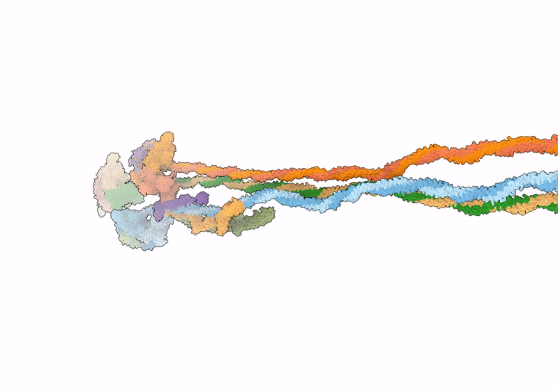
PDB ID:
8Q6T
Keywords:
Title:
Helical reconstruction of the relaxed thick filament from FIB milled left ventricular mouse myofibrils
Biological Source:
Source Organism:
Mus musculus (Taxon ID: 10090)
Method Details:
Experimental Method:
Resolution:
18.00 Å
Aggregation State:
CELL
Reconstruction Method:
SUBTOMOGRAM AVERAGING
