8Q5U image
Deposition Date 2023-08-09
Release Date 2024-04-17
Last Version Date 2024-10-09
Entry Detail
PDB ID:
8Q5U
Keywords:
Title:
Endoglycosidase S2 in complex with IgG1 Fc
Biological Source:
Source Organism:
Host Organism:
Method Details:
Experimental Method:
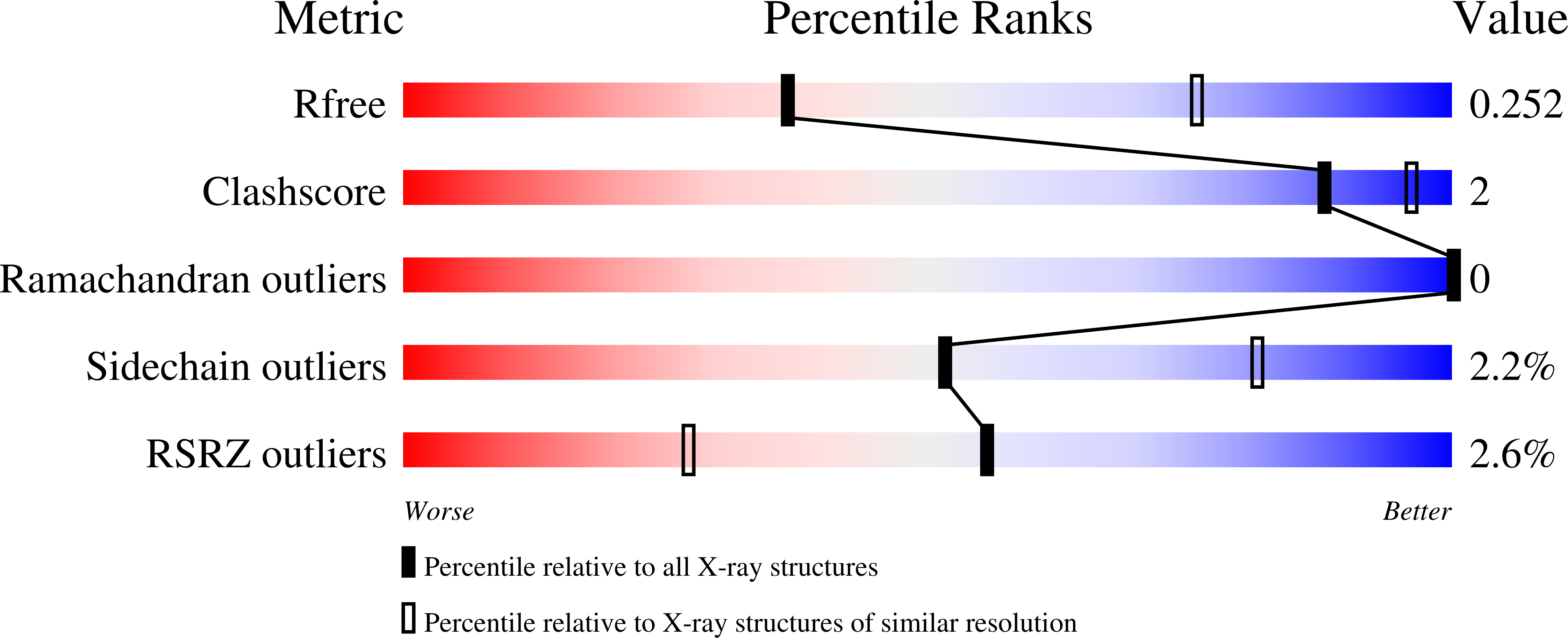
Resolution:
3.00 Å
R-Value Free:
0.25
R-Value Work:
0.21
Space Group:
P 43 21 2
Macromolecular Entities
Structures with similar UniProt ID
Protein Blast
Polymer Type:polypeptide(L)
Molecule:Uncharacterized protein DKFZp686C11235
Mutagens:L234C E382A
Chain IDs:A, C, F (auth: B)
Chain Length:227
Number of Molecules:3
Biological Source:Homo sapiens
Structures with similar UniProt ID
Protein Blast
Polymer Type:polypeptide(L)
Molecule:Endo-beta-N-acetylglucosaminidase
Mutagens:D184A E186L
Chain IDs:B (auth: D), D (auth: E), E (auth: F)
Chain Length:816
Number of Molecules:3
Biological Source:Streptococcus pyogenes
Primary Citation
The IgG-specific endoglycosidases EndoS and EndoS2 are distinguished by conformation and antibody recognition.
J.Biol.Chem. 300 107245 107245 (2024)
PMID: 38569940 DOI: 10.1016/j.jbc.2024.107245

Abstact

The IgG-specific endoglycosidases EndoS and EndoS2 from Streptococcus pyogenes can remove conserved N-linked glycans present on the Fc region of host antibodies to inhibit Fc-mediated effector functions. These enzymes are therefore being investigated as therapeutics for suppressing unwanted immune activation, and have additional application as tools for antibody glycan remodeling. EndoS and EndoS2 differ in Fc glycan substrate specificity due to structural differences within their catalytic glycosyl hydrolase domains. However, a chimeric EndoS enzyme with a substituted glycosyl hydrolase from EndoS2 loses catalytic activity, despite high structural homology between the two enzymes, indicating either mechanistic divergence of EndoS and EndoS2, or improperly-formed domain interfaces in the chimeric enzyme. Here, we present the crystal structure of the EndoS2-IgG1 Fc complex determined to 3.0 Å resolution. Comparison of complexed and unliganded EndoS2 reveals relative reorientation of the glycosyl hydrolase, leucine-rich repeat and hybrid immunoglobulin domains. The conformation of the complexed EndoS2 enzyme is also different when compared to the earlier EndoS-IgG1 Fc complex, and results in distinct contact surfaces between the two enzymes and their Fc substrate. These findings indicate mechanistic divergence of EndoS2 and EndoS. It will be important to consider these differences in the design of IgG-specific enzymes, developed to enable customizable antibody glycosylation.

Legend

Protein

Chemical

Disease

Primary Citation of related structures