
Deposition Date
2023-08-02
Release Date
2023-11-01
Last Version Date
2023-11-15
Entry Detail
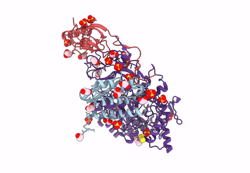
PDB ID:
8Q2E
Keywords:
Title:
The 1.68-A X-ray crystal structure of Sporosarcina pasteurii urease inhibited by thiram and bound to dimethylditiocarbamate
Biological Source:
Source Organism(s):
Sporosarcina pasteurii (Taxon ID: 1474)
Method Details:
Experimental Method:
Resolution:
1.68 Å
R-Value Free:
0.16
R-Value Work:
0.13
R-Value Observed:
0.13
Space Group:
P 63 2 2
