Deposition Date
2023-08-01
Release Date
2024-07-10
Last Version Date
2024-07-10
Entry Detail
PDB ID:
8Q2B
Keywords:
Title:
E. coli Adenylate Kinase variant D158A (AK D158A) showing significant changes to the stacking of catalytic arginine side chains
Biological Source:
Source Organism(s):
Escherichia coli (Taxon ID: 562)
Expression System(s):
Method Details:
Experimental Method:
Resolution:
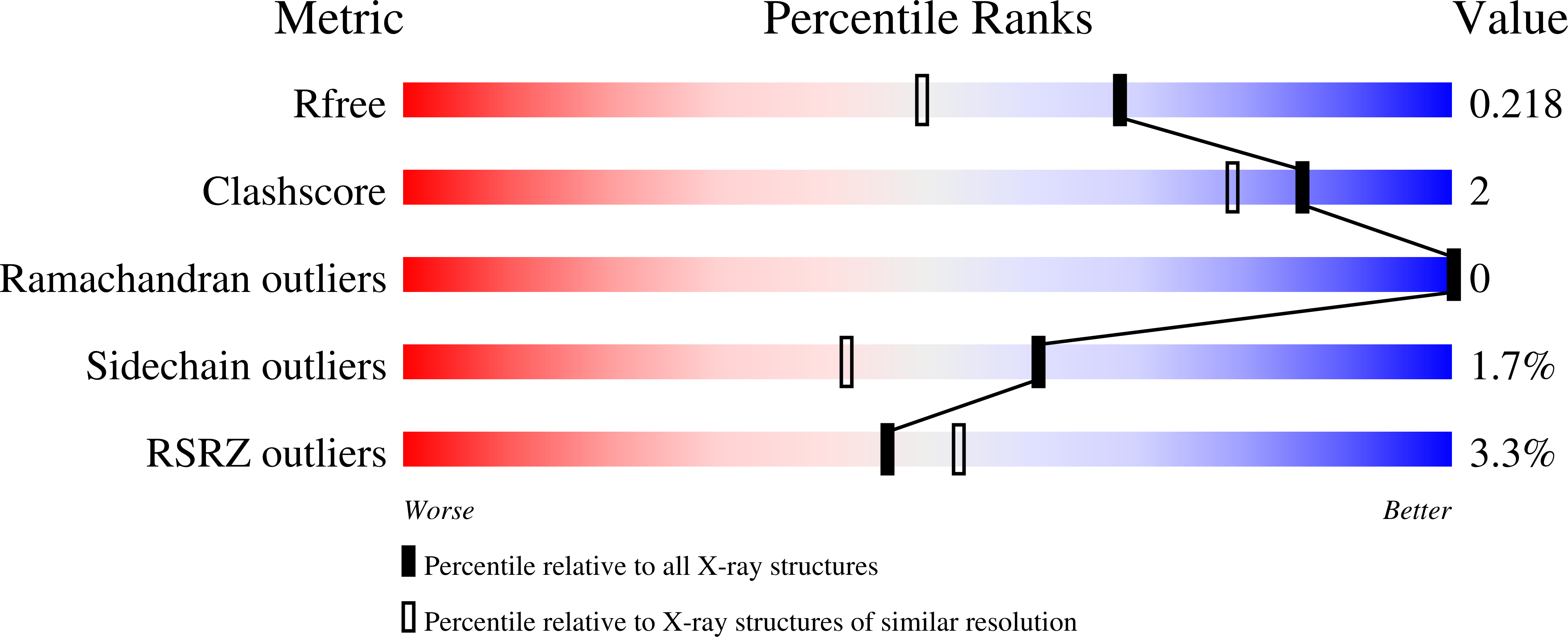
1.76 Å
R-Value Free:
0.22
R-Value Work:
0.18
R-Value Observed:
0.18
Space Group:
P 1 21 1