8PX0 image
Deposition Date 2023-07-22
Release Date 2023-10-25
Last Version Date 2024-11-06
Entry Detail
PDB ID:
8PX0
Title:
Structure of ribonuclease A, solved at wavelength 2.75 A
Biological Source:
Source Organism:
Bos taurus (Taxon ID: 9913)
Host Organism:
Method Details:
Experimental Method:
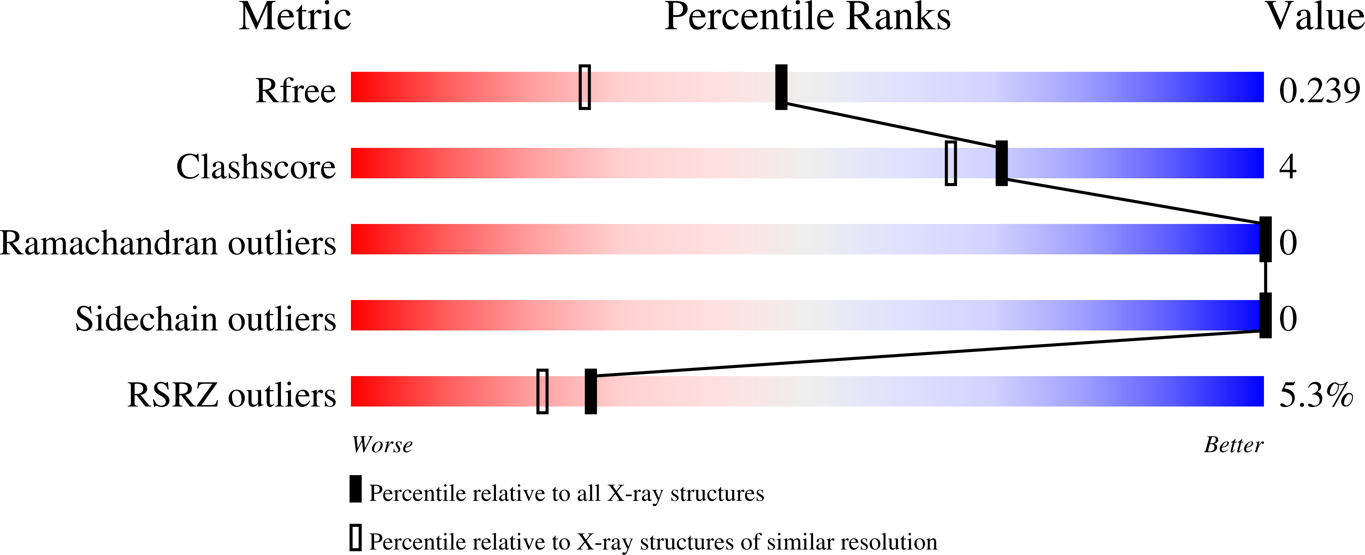
Resolution:
1.80 Å
R-Value Free:
0.23
R-Value Work:
0.20
R-Value Observed:
0.20
Space Group:
C 1 2 1
Macromolecular Entities
Structures with similar UniProt ID
Protein Blast
Polymer Type:polypeptide(L)
Molecule:Ribonuclease pancreatic
Gene (Uniprot):RNASE1
Chain IDs:A, B
Chain Length:124
Number of Molecules:2
Biological Source:Bos taurus
Ligand Molecules
Primary Citation

Abstact

Despite recent advances in cryo-electron microscopy and artificial intelligence-based model predictions, a significant fraction of structure determinations by macromolecular crystallography still requires experimental phasing, usually by means of single-wavelength anomalous diffraction (SAD) techniques. Most synchrotron beamlines provide highly brilliant beams of X-rays of between 0.7 and 2 Å wavelength. Use of longer wavelengths to access the absorption edges of biologically important lighter atoms such as calcium, potassium, chlorine, sulfur and phosphorus for native-SAD phasing is attractive but technically highly challenging. The long-wavelength beamline I23 at Diamond Light Source overcomes these limitations and extends the accessible wavelength range to λ = 5.9 Å. Here we report 22 macromolecular structures solved in this extended wavelength range, using anomalous scattering from a range of elements which demonstrate the routine feasibility of lighter atom phasing. We suggest that, in light of its advantages, long-wavelength crystallography is a compelling option for experimental phasing.

Legend

Protein

Chemical

Disease

Primary Citation of related structures
Feedback Form
Name
Email
Institute
Feedback