8PWN image
Entry Detail
PDB ID:
8PWN
Title:
Structure of A2A adenosine receptor A2AR-StaR2-bRIL, solved at wavelength 2.75 A
Biological Source:
Source Organism:
Host Organism:
PDB Version:
Deposition Date:
2023-07-20
Release Date:
2023-10-25
Method Details:
Experimental Method:
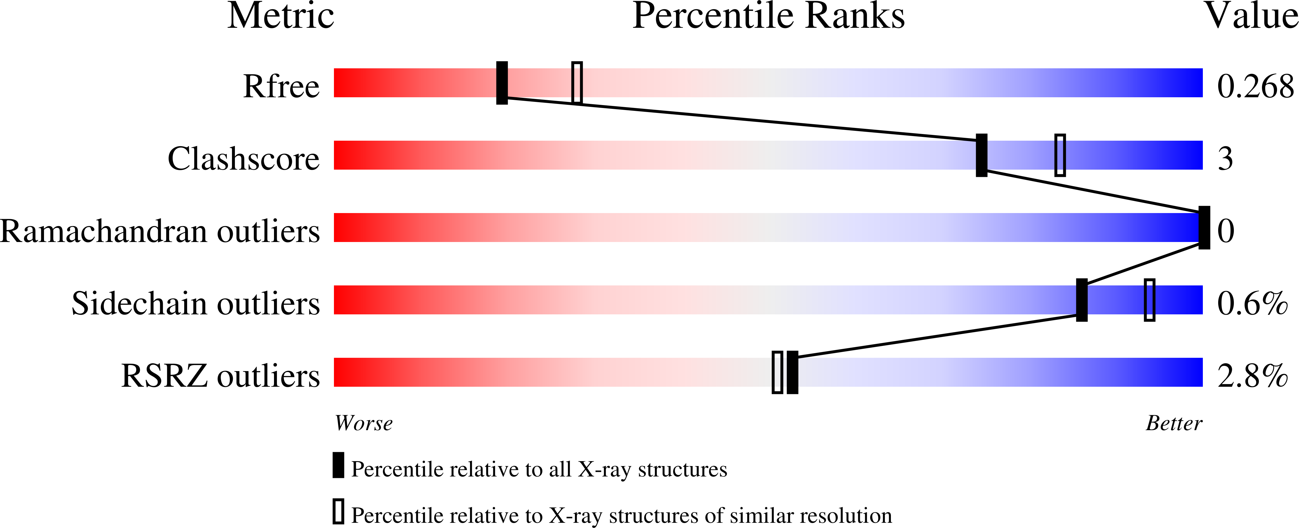
Resolution:
2.40 Å
R-Value Free:
0.26
R-Value Work:
0.22
R-Value Observed:
0.22
Space Group:
C 2 2 21
Macromolecular Entities
Polymer Type:polypeptide(L)
Description:Adenosine receptor A2a,Soluble cytochrome b562
Chain IDs:A
Chain Length:433
Number of Molecules:1
Biological Source:Homo sapiens, Escherichia coli
Primary Citation

Abstact

Despite recent advances in cryo-electron microscopy and artificial intelligence-based model predictions, a significant fraction of structure determinations by macromolecular crystallography still requires experimental phasing, usually by means of single-wavelength anomalous diffraction (SAD) techniques. Most synchrotron beamlines provide highly brilliant beams of X-rays of between 0.7 and 2 Å wavelength. Use of longer wavelengths to access the absorption edges of biologically important lighter atoms such as calcium, potassium, chlorine, sulfur and phosphorus for native-SAD phasing is attractive but technically highly challenging. The long-wavelength beamline I23 at Diamond Light Source overcomes these limitations and extends the accessible wavelength range to λ = 5.9 Å. Here we report 22 macromolecular structures solved in this extended wavelength range, using anomalous scattering from a range of elements which demonstrate the routine feasibility of lighter atom phasing. We suggest that, in light of its advantages, long-wavelength crystallography is a compelling option for experimental phasing.

Legend

Protein

Chemical

Disease

Primary Citation of related structures