Deposition Date
2023-07-05
Release Date
2024-09-18
Last Version Date
2024-09-18
Entry Detail
PDB ID:
8POX
Keywords:
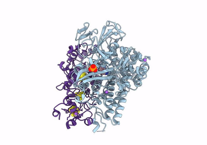
Title:
Crystal Structure of the C19G variant of the membrane-bound [NiFe]-Hydrogenase from Cupriavidus necator in the H2-reduced state at 1.6 A Resolution.
Biological Source:
Source Organism(s):
Cupriavidus necator H16 (Taxon ID: 381666)
Expression System(s):
Method Details:
Experimental Method:
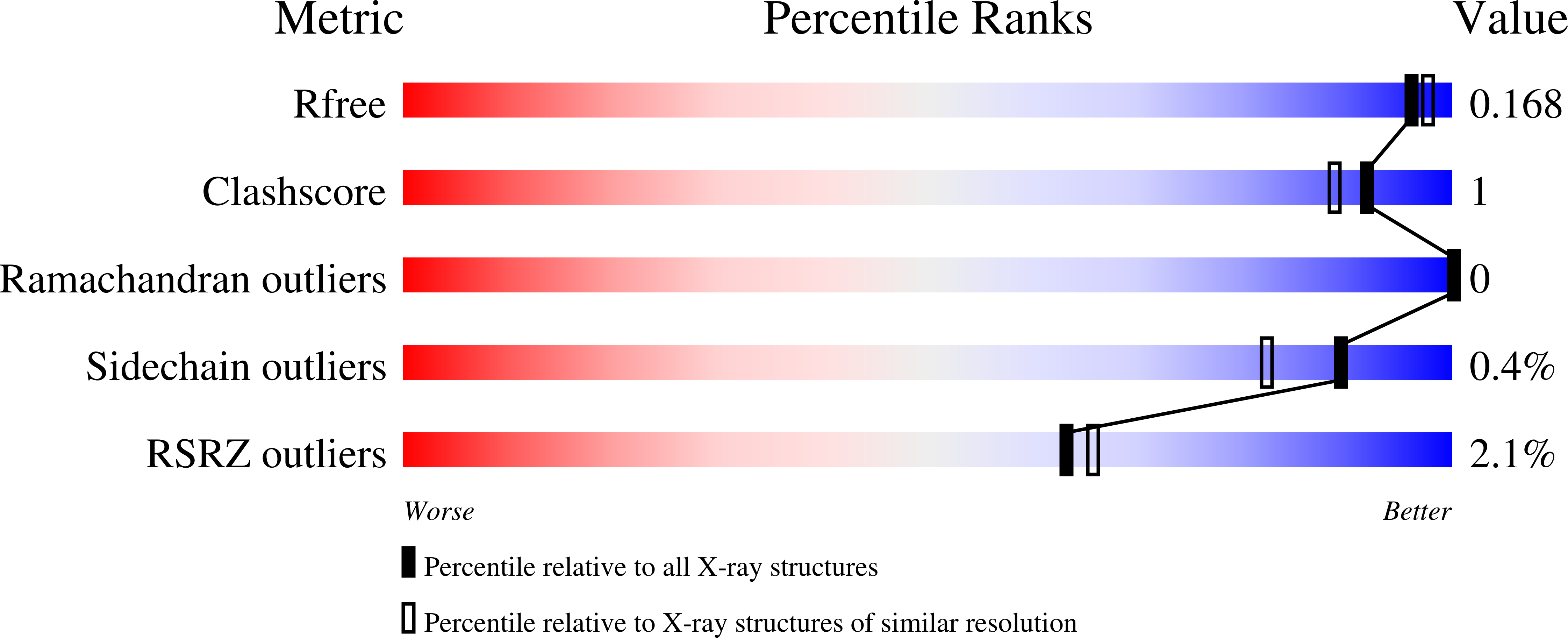
Resolution:
1.60 Å
R-Value Free:
0.15
R-Value Work:
0.12
R-Value Observed:
0.12
Space Group:
P 21 21 21