8P8E image
Deposition Date 2023-06-01
Release Date 2024-05-08
Last Version Date 2024-08-21
Entry Detail
PDB ID:
8P8E
Keywords:
Title:
Crystal structure of endolysin gp46 from Pseudomonas aeruginosa bacteriophage vB_PaeM_KTN6
Biological Source:
Source Organism:
Host Organism:
Method Details:
Experimental Method:
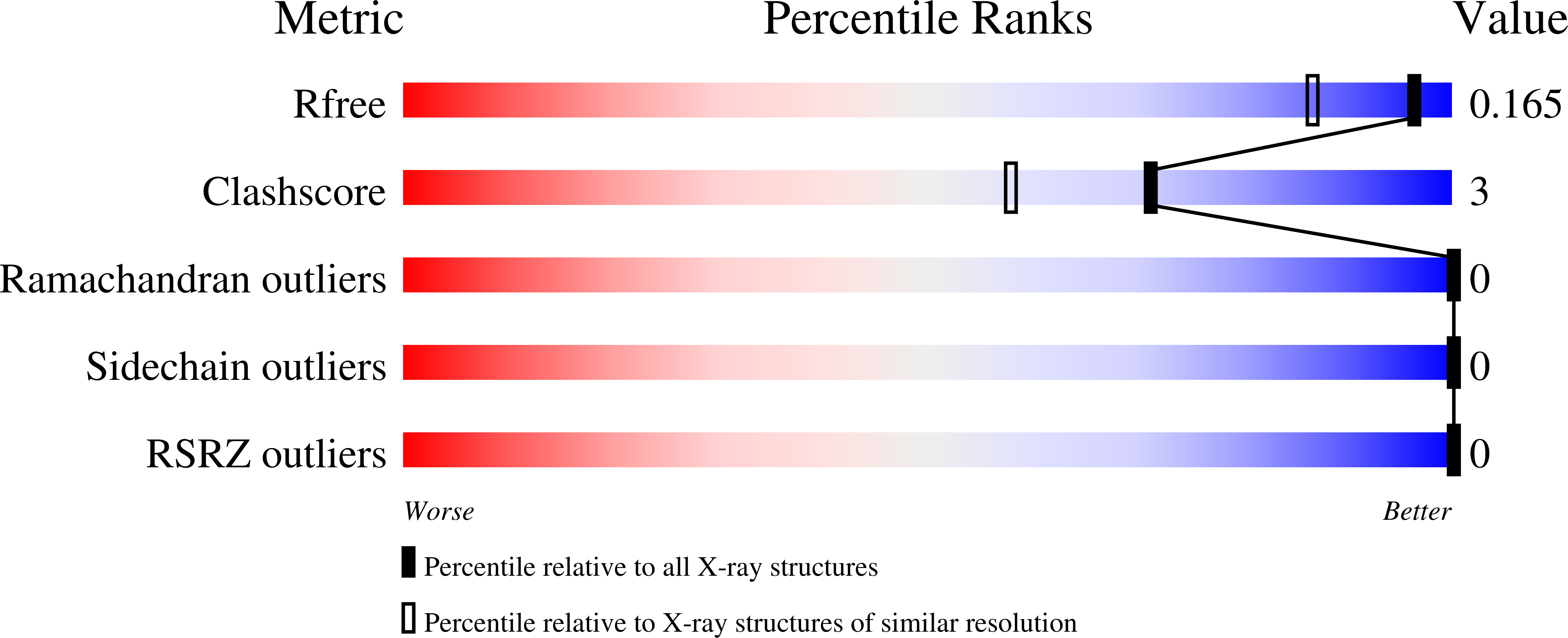
Resolution:
1.39 Å
R-Value Free:
0.16
R-Value Work:
0.12
Space Group:
P 21 21 21
Macromolecular Entities
Structures with similar UniProt ID
Protein Blast
Polymer Type:polypeptide(L)
Molecule:Glycoside hydrolase family protein
Gene (Uniprot):IDBGMNHM_00003
Chain IDs:A (auth: AAA)
Chain Length:220
Number of Molecules:1
Biological Source:Bacteriophage sp.
Primary Citation

Abstact

BACKGROUND Endolysins are phage-encoded lytic enzymes that degrade bacterial peptidoglycan at the end of phage lytic cycles to release new phage particles. These enzymes are being explored as an alternative to small-molecule antibiotics. METHODS The crystal structure of KTN6 Gp46 was determined and compared with a ColabFold model. Cleavage specificity was examined using a peptidoglycan digest and reversed-phase high-performance liquid chromatography coupled to mass spectrometry (HPLC/MS). RESULTS The structure of KTN6 Gp46 could be determined at 1.4 Å resolution, and key differences in loops of the putative peptidoglycan binding domain were identified in comparison with its closest known homologue, the endolysin of phage SPN1S. Reversed-phase HPLC/MS analysis of the reaction products following peptidoglycan digestion confirmed the muramidase activity of Gp46, consistent with structural predictions. CONCLUSION These insights into the structure and function of endolysins further expand the toolbox for endolysin engineering and explore their potential in enzyme-based antibacterial design strategies.

Legend

Protein

Chemical

Disease

Primary Citation of related structures