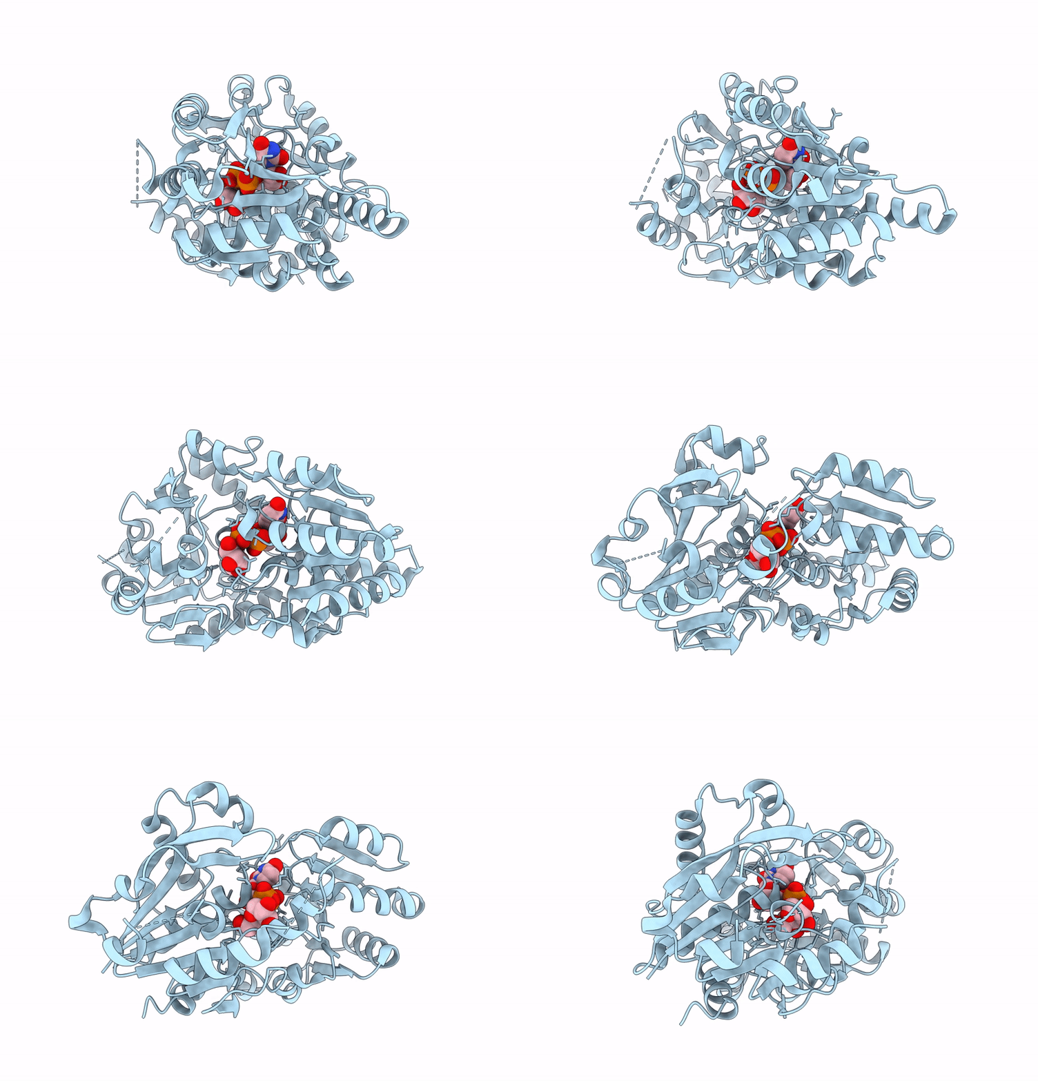
8P6S image
Deposition Date 2023-05-29
Release Date 2024-12-11
Last Version Date 2025-06-25
Entry Detail
PDB ID:
8P6S
Keywords:
Title:
Arabidopsis thaliana DGD2 in complex with UDP-galactose
Biological Source:
Source Organism:
Host Organism:
Method Details:
Experimental Method:
Resolution:
2.10 Å
R-Value Free:
0.25
R-Value Work:
0.20
R-Value Observed:
0.20
Space Group:
P 41 21 2
Macromolecular Entities
Polymer Type:polypeptide(L)
Molecule:Digalactosyldiacylglycerol synthase 2, chloroplastic
Gene (Uniprot):DGD2
Chain IDs:A
Chain Length:412
Number of Molecules:1
Biological Source:Arabidopsis thaliana
Ligand Molecules
Primary Citation
Structure and membrane interactions of Arabidopsis thaliana DGD2, a glycosyltransferase in the chloroplast membrane.
J.Biol.Chem. 301 108431 108431 (2025)
PMID: 40120685 DOI: 10.1016/j.jbc.2025.108431

Abstact

Galactolipids are characteristic lipids of the photosynthesis membranes of higher plants and cyanobacteria. Due to their close relationship to the stability of the photosystem protein complexes, the biogenesis of galactolipids has been intensively studied on the genetic and molecular levels. There are two major types of galactolipids in chloroplastic membranes: monogalactosyldiacylglycerol and digalactosyldiacylglycerol (DGDG). Under phosphate-limiting conditions, the amount of DGDG increases dramatically to allow for phosphate salvage from phospholipids. In Arabidopsis thaliana, the membrane-associated glycosyltransferase digalactosyldiacylglycerol synthase 2 (atDGD2) is highly responsive to phosphate starvation and is significantly upregulated during such conditions. The lipid galactosylation reactions are also fundamentally interesting as they require a catalyst that is capable of bringing a hydrophilic and lipophilic substrate together at the solution-membrane phase border. Here, we present the X-ray crystal structure of atDGD2, which is the first reported DGDG synthase structure. AtDGD2 is most structurally similar to functionally unrelated GT-B enzymes. Interestingly, in spite of significant donor substrate binding differences, we identified four amino acids (Gly22, His151, Lys243, and Glu321, atDGD2 numbering) which were entirely conserved between the structurally similar enzymes. We also investigated the membrane interaction kinetics and membrane anchoring mechanism of atDGD2. This demonstrated that atDGD2 is membrane-bound but also showed that membrane binding is highly dynamic. Furthermore, our structural information in context of previous biophysical studies highlights regions of the enzyme exhibiting a high degree of structural plasticity, which we propose to be important for allowing atDGD2 to quickly adapt its activity based on the membrane lipid environment.

Legend

Protein

Chemical

Disease

Primary Citation of related structures