Deposition Date
2023-05-10
Release Date
2023-11-29
Last Version Date
2024-10-16
Entry Detail
PDB ID:
8P0L
Keywords:
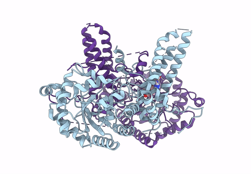
Title:
Crystal structure of human O-GlcNAcase in complex with an S-linked CKII peptide
Biological Source:
Source Organism(s):
Homo sapiens (Taxon ID: 9606)
Expression System(s):
Method Details:
Experimental Method:
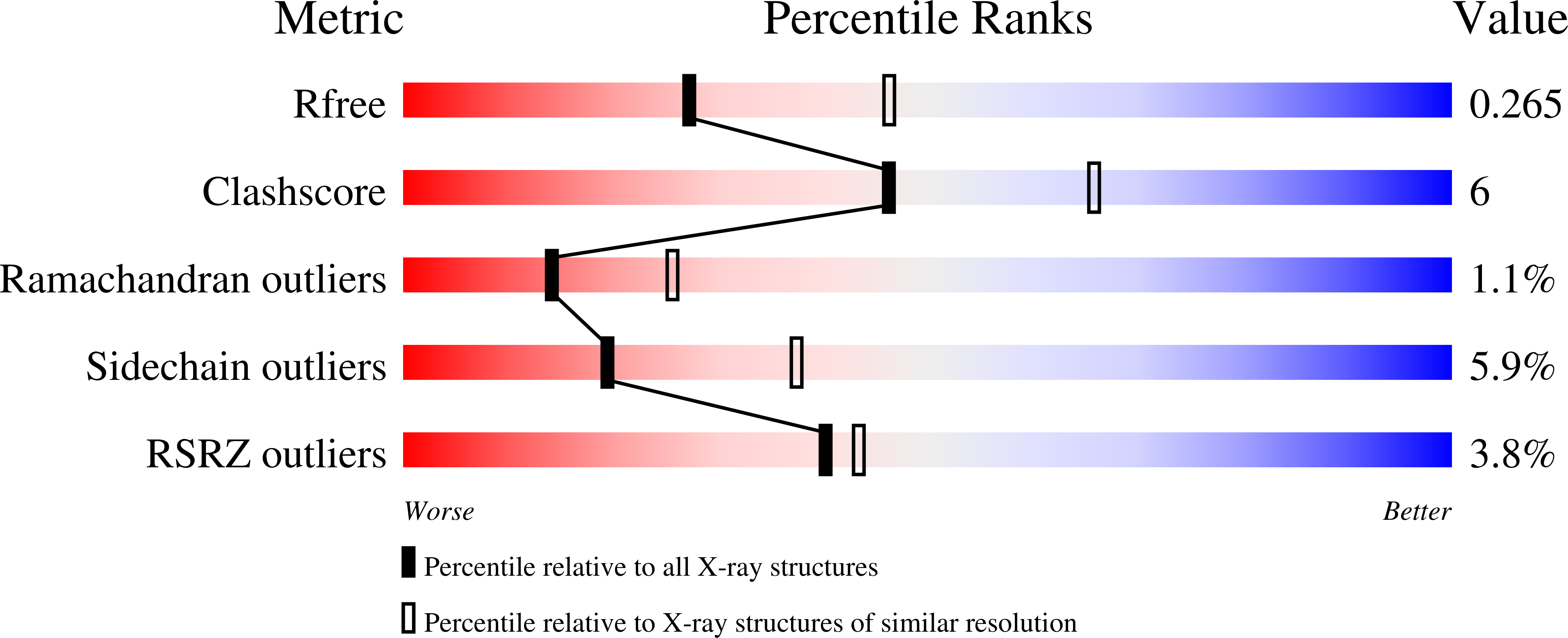
Resolution:
2.50 Å
R-Value Free:
0.27
R-Value Work:
0.21
Space Group:
P 43 21 2