8OSC image
Entry Detail
PDB ID:
8OSC
Keywords:
Title:
Structure of Homo sapiens 2'-deoxynucleoside 5'-phosphate N-hydrolase 1 (DNPH1) bound to deoxyuridine 5'- monophosphate
Biological Source:
Source Organism:
Host Organism:
PDB Version:
Deposition Date:
2023-04-18
Release Date:
2023-08-30
Method Details:
Experimental Method:
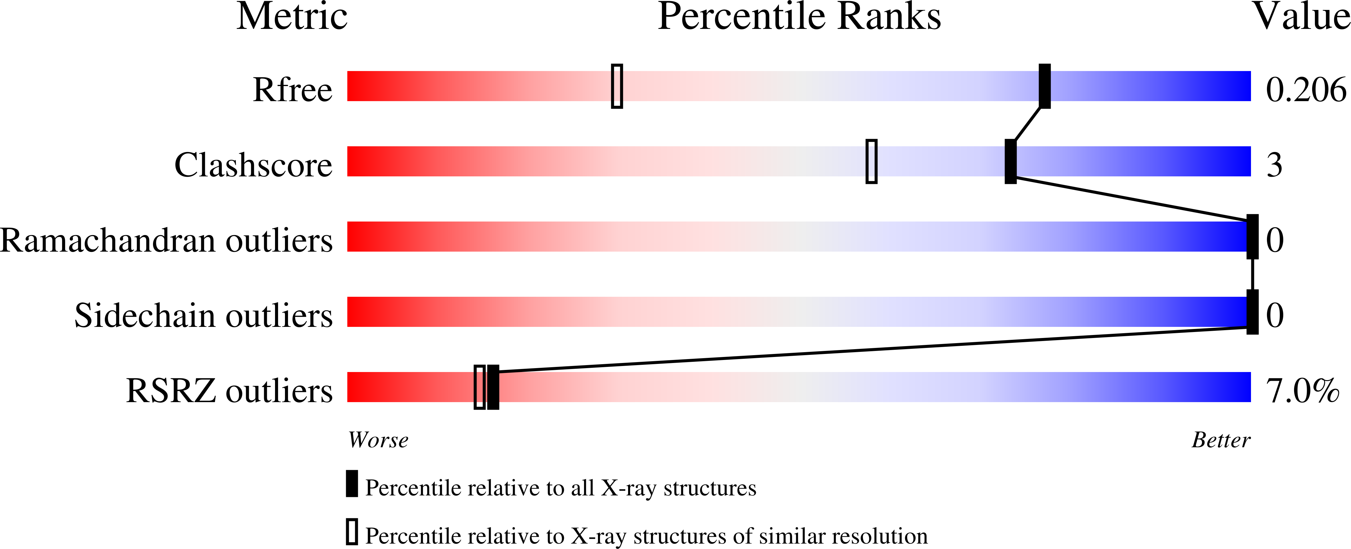
Resolution:
1.42 Å
R-Value Free:
0.21
R-Value Work:
0.19
R-Value Observed:
0.19
Space Group:
C 1 2 1
Macromolecular Entities
Polymer Type:polypeptide(L)
Description:2'-deoxynucleoside 5'-phosphate N-hydrolase 1
Chain IDs:A, B
Chain Length:145
Number of Molecules:2
Biological Source:Homo sapiens
Ligand Molecules
Primary Citation
Human 2'-Deoxynucleoside 5'-Phosphate N -Hydrolase 1: Mechanism of 2'-Deoxyuridine 5'-Monophosphate Hydrolysis.
Biochemistry 62 2658 2668 (2023)
PMID: 37582341 DOI: 10.1021/acs.biochem.3c00369

Abstact

The enzyme 2'-deoxynucleoside 5'-phosphate N-hydrolase 1 (DNPH1) catalyzes the N-ribosidic bond cleavage of 5-hydroxymethyl-2'-deoxyuridine 5'-monophosphate to generate 2-deoxyribose 5-phosphate and 5-hydroxymethyluracil. DNPH1 accepts other 2'-deoxynucleoside 5'-monophosphates as slow-reacting substrates. DNPH1 inhibition is a promising strategy to overcome resistance to and potentiate anticancer poly(ADP-ribose) polymerase inhibitors. We solved the crystal structure of unliganded human DNPH1 and took advantage of the slow reactivity of 2'-deoxyuridine 5'-monophosphate (dUMP) as a substrate to obtain a crystal structure of the DNPH1:dUMP Michaelis complex. In both structures, the carboxylate group of the catalytic Glu residue, proposed to act as a nucleophile in covalent catalysis, forms an apparent low-barrier hydrogen bond with the hydroxyl group of a conserved Tyr residue. The crystal structures are supported by functional data, with liquid chromatography-mass spectrometry analysis showing that DNPH1 incubation with dUMP leads to slow yet complete hydrolysis of the substrate. A direct UV-vis absorbance-based assay allowed characterization of DNPH1 kinetics at low dUMP concentrations. A bell-shaped pH-rate profile indicated that acid-base catalysis is operational and that for maximum kcat/KM, two groups with an average pKa of 6.4 must be deprotonated, while two groups with an average pKa of 8.2 must be protonated. A modestly inverse solvent viscosity effect rules out diffusional processes involved in dUMP binding to and possibly uracil release from the enzyme as rate limiting to kcat/KM. Solvent deuterium isotope effects on kcat/KM and kcat were inverse and unity, respectively. A reaction mechanism for dUMP hydrolysis is proposed.

Legend

Protein

Chemical

Disease

Primary Citation of related structures