8OR7 image
Entry Detail
PDB ID:
8OR7
Keywords:
Title:
Structure of a far-red induced allophycocyanin from Chroococcidiopsis thermalis sp. PCC 7203
Biological Source:
Host Organism:
PDB Version:
Deposition Date:
2023-04-13
Release Date:
2024-03-20
Method Details:
Experimental Method:
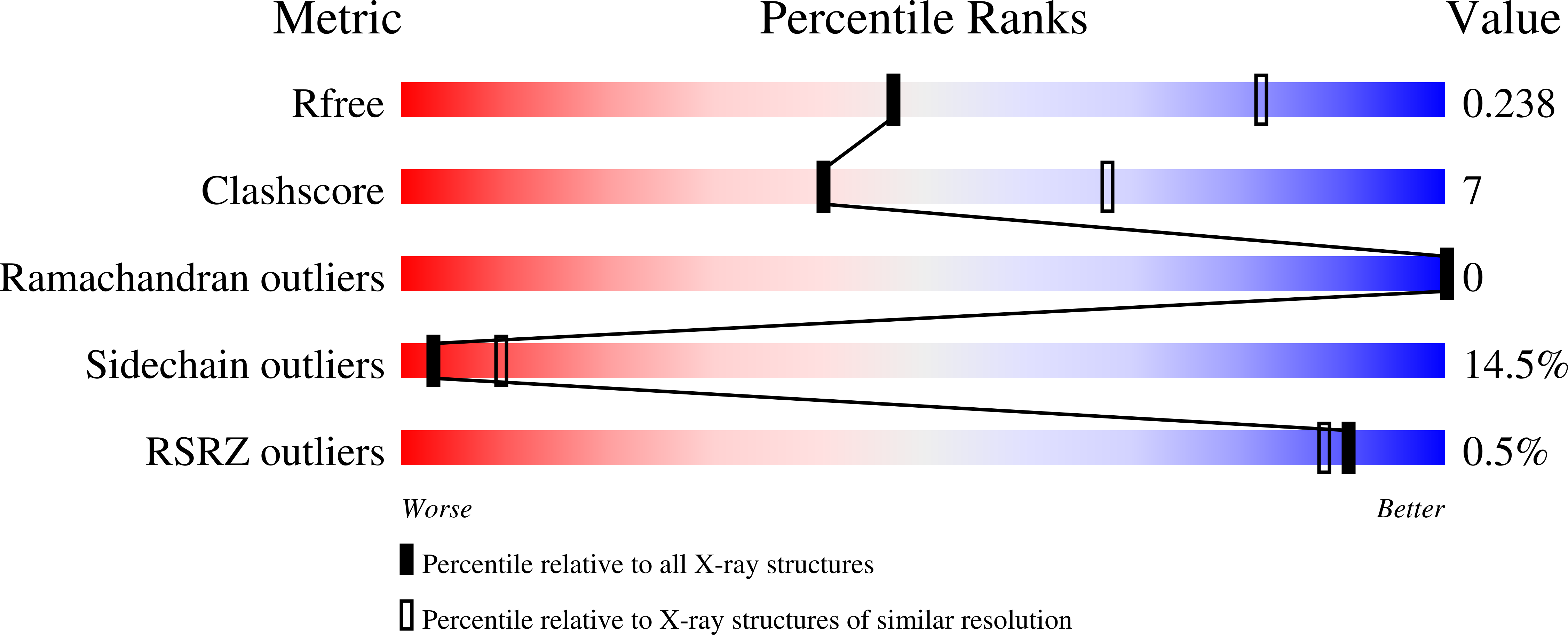
Resolution:
2.80 Å
R-Value Free:
0.24
R-Value Work:
0.17
R-Value Observed:
0.18
Space Group:
P 63
Macromolecular Entities
Polymer Type:polypeptide(L)
Description:Phycocyanin
Chain IDs:C (auth: A), D (auth: C)
Chain Length:159
Number of Molecules:2
Biological Source:Chroococcidiopsis thermalis
Polymer Type:polypeptide(L)
Description:Allophycocyanin beta subunit apoprotein
Chain IDs:A (auth: B), B (auth: D)
Chain Length:169
Number of Molecules:2
Biological Source:Chroococcidiopsis thermalis
Primary Citation
Crystallographic and biochemical analyses of a far-red allophycocyanin to address the mechanism of the super-red-shift.
Photosynth.Res. 162 171 185 (2024)
PMID: 38182842 DOI: 10.1007/s11120-023-01066-2

Abstact

Far-red absorbing allophycocyanins (APC), identified in cyanobacteria capable of FRL photoacclimation (FaRLiP) and low-light photoacclimation (LoLiP), absorb far-red light, functioning in energy transfer as light-harvesting proteins. We report an optimized method to obtain high purity far-red absorbing allophycocyanin B, AP-B2, of Chroococcidiopsis thermalis sp. PCC7203 by synthesis in Escherichia coli and an improved purification protocol. The crystal structure of the trimer, (PCB-ApcD5/PCB-ApcB2)3, has been resolved to 2.8 Å. The main difference to conventional APCs absorbing in the 650-670 nm range is a largely flat chromophore with the co-planarity extending, in particular, from rings BCD to ring A. This effectively extends the conjugation system of PCB and contributes to the super-red-shifted absorption of the α-subunit (λmax = 697 nm). On complexation with the β-subunit, it is even further red-shifted (λmax, absorption = 707 nm, λmax, emission = 721 nm). The relevance of ring A for this shift is supported by mutagenesis data. A variant of the α-subunit, I123M, has been generated that shows an intense FR-band already in the absence of the β-subunit, a possible model is discussed. Two additional mechanisms are known to red-shift the chromophore spectrum: lactam-lactim tautomerism and deprotonation of the chromophore that both mechanisms appear inconsistent with our data, leaving this question unresolved.

Legend

Protein

Chemical

Disease

Primary Citation of related structures