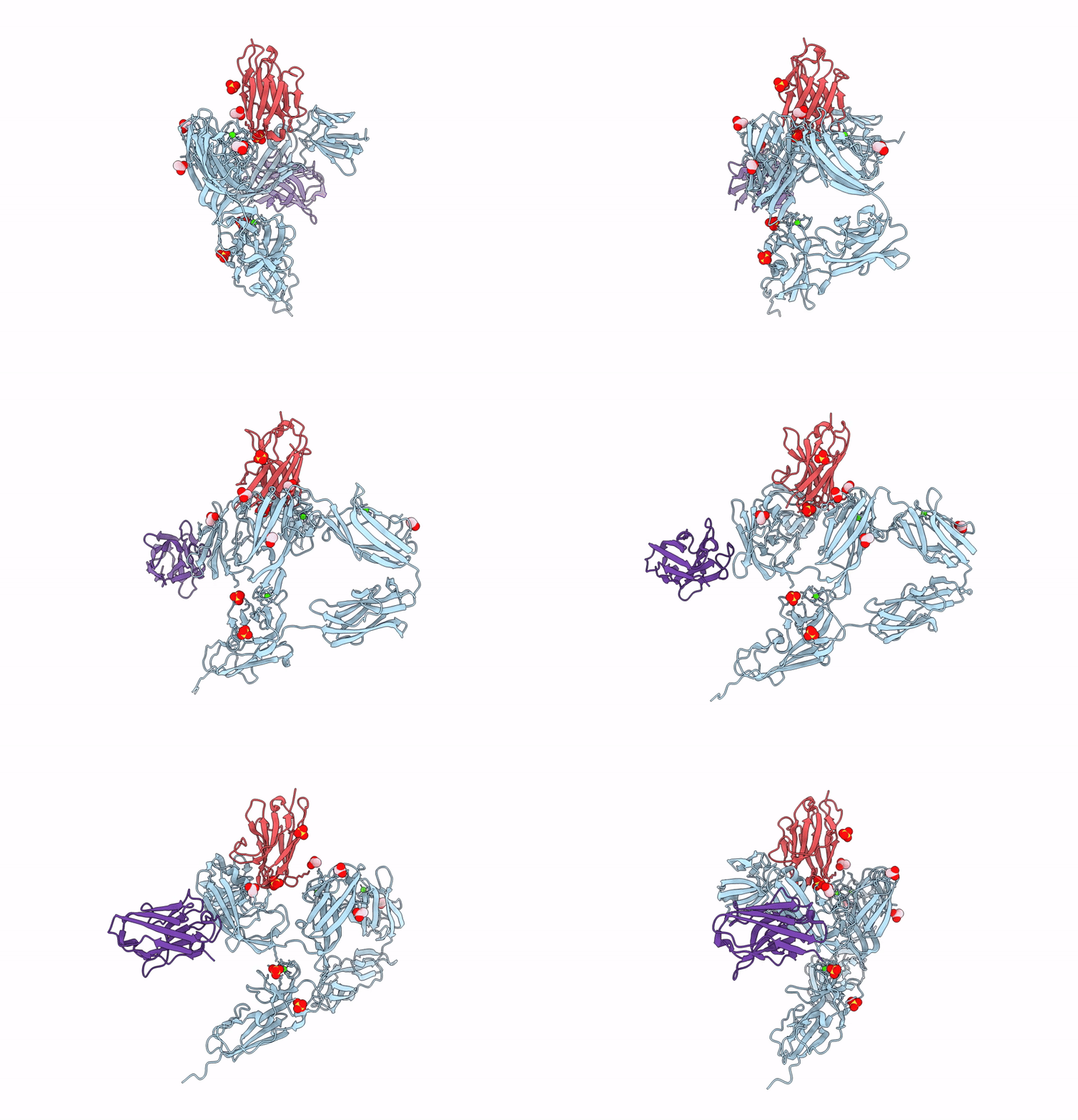
8OPR image
Entry Detail
PDB ID:
8OPR
Title:
Structure of the EA1 surface layer of Bacillus anthracis
Biological Source:
Source Organism:
PDB Version:
Deposition Date:
2023-04-07
Release Date:
2023-11-08
Method Details:
Experimental Method:
Resolution:
1.81 Å
R-Value Free:
0.24
R-Value Work:
0.20
R-Value Observed:
0.20
Space Group:
P 1
Macromolecular Entities
Polymer Type:polypeptide(L)
Description:S-layer protein EA1
Chain IDs:A
Chain Length:665
Number of Molecules:1
Biological Source:Bacillus anthracis
Polymer Type:polypeptide(L)
Description:Nanobody 643
Chain IDs:B
Chain Length:127
Number of Molecules:1
Biological Source:Lama glama
Polymer Type:polypeptide(L)
Description:Nanobody 632
Chain IDs:C
Chain Length:133
Number of Molecules:1
Biological Source:Lama glama
Primary Citation
Structure and function of the EA1 surface layer of Bacillus anthracis.
Nat Commun 14 7051 7051 (2023)
PMID: 37923757 DOI: 10.1038/s41467-023-42826-x

Abstact

The Gram-positive spore-forming bacterium Bacillus anthracis is the causative agent of anthrax, a deadly disease mostly affecting wildlife and livestock, as well as representing a bioterrorism threat. Its cell surface is covered by the mutually exclusive S-layers Sap and EA1, found in early and late growth phases, respectively. Here we report the nanobody-based structural characterization of EA1 and its native lattice contacts. The EA1 assembly domain consists of 6 immunoglobulin-like domains, where three calcium-binding sites structure interdomain contacts that allow monomers to adopt their assembly-competent conformation. Nanobody-induced depolymerization of EA1 S-layers results in surface defects, membrane blebbing and cell lysis under hypotonic conditions, indicating that S-layers provide additional mechanical stability to the cell wall. Taken together, we report a complete model of the EA1 S-layer and present a set of nanobodies that may have therapeutic potential against Bacillus anthracis.

Legend

Protein

Chemical

Disease

Primary Citation of related structures