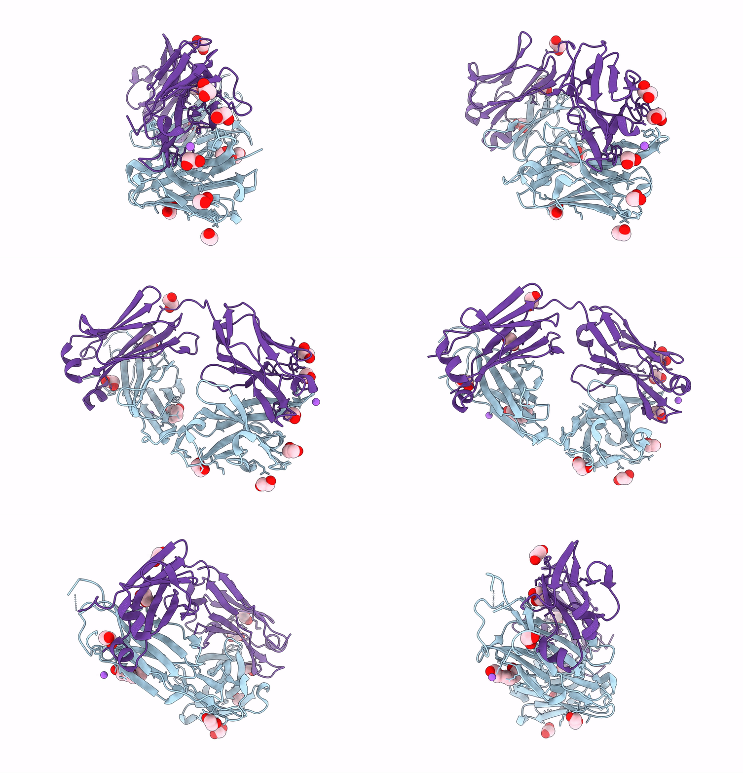
8OJU image
Entry Detail
PDB ID:
8OJU
Keywords:
Title:
Crystal structure of the human IgD Fab - structure Fab3
Biological Source:
Source Organism:
Host Organism:
PDB Version:
Deposition Date:
2023-03-24
Release Date:
2023-06-14
Method Details:
Experimental Method:
Resolution:
1.45 Å
R-Value Free:
0.18
R-Value Work:
0.15
R-Value Observed:
0.15
Space Group:
P 1 21 1
Macromolecular Entities
Polymer Type:polypeptide(L)
Description:Human IgD Fab heavy chain
Chain IDs:B (auth: H)
Chain Length:227
Number of Molecules:1
Biological Source:Homo sapiens
Polymer Type:polypeptide(L)
Description:Human IgD Fab light chain
Chain IDs:A (auth: L)
Chain Length:217
Number of Molecules:1
Biological Source:Homo sapiens
Primary Citation
Crystal structures of the human IgD Fab reveal insights into C H 1 domain diversity.
Mol.Immunol. 159 28 37 (2023)
PMID: 37267832 DOI: 10.1016/j.molimm.2023.05.006

Abstact

Antibodies of the IgD isotype remain the least well characterized of the mammalian immunoglobulin isotypes. Here we report three-dimensional structures for the Fab region of IgD, based on four different crystal structures, at resolutions of 1.45-2.75 Å. These IgD Fab crystals provide the first high-resolution views of the unique Cδ1 domain. Structural comparisons identify regions of conformational diversity within the Cδ1 domain, as well as among the homologous domains of Cα1, Cγ1 and Cμ1. The IgD Fab structure also possesses a unique conformation of the upper hinge region, which may contribute to the overall disposition of the very long linker sequence between the Fab and Fc regions found in human IgD. Structural similarities observed between IgD and IgG, and differences with IgA and IgM, are consistent with predicted evolutionary relationships for the mammalian antibody isotypes.

Legend

Protein

Chemical

Disease

Primary Citation of related structures