
Deposition Date
1988-01-04
Release Date
1989-07-12
Last Version Date
2024-10-16
Entry Detail
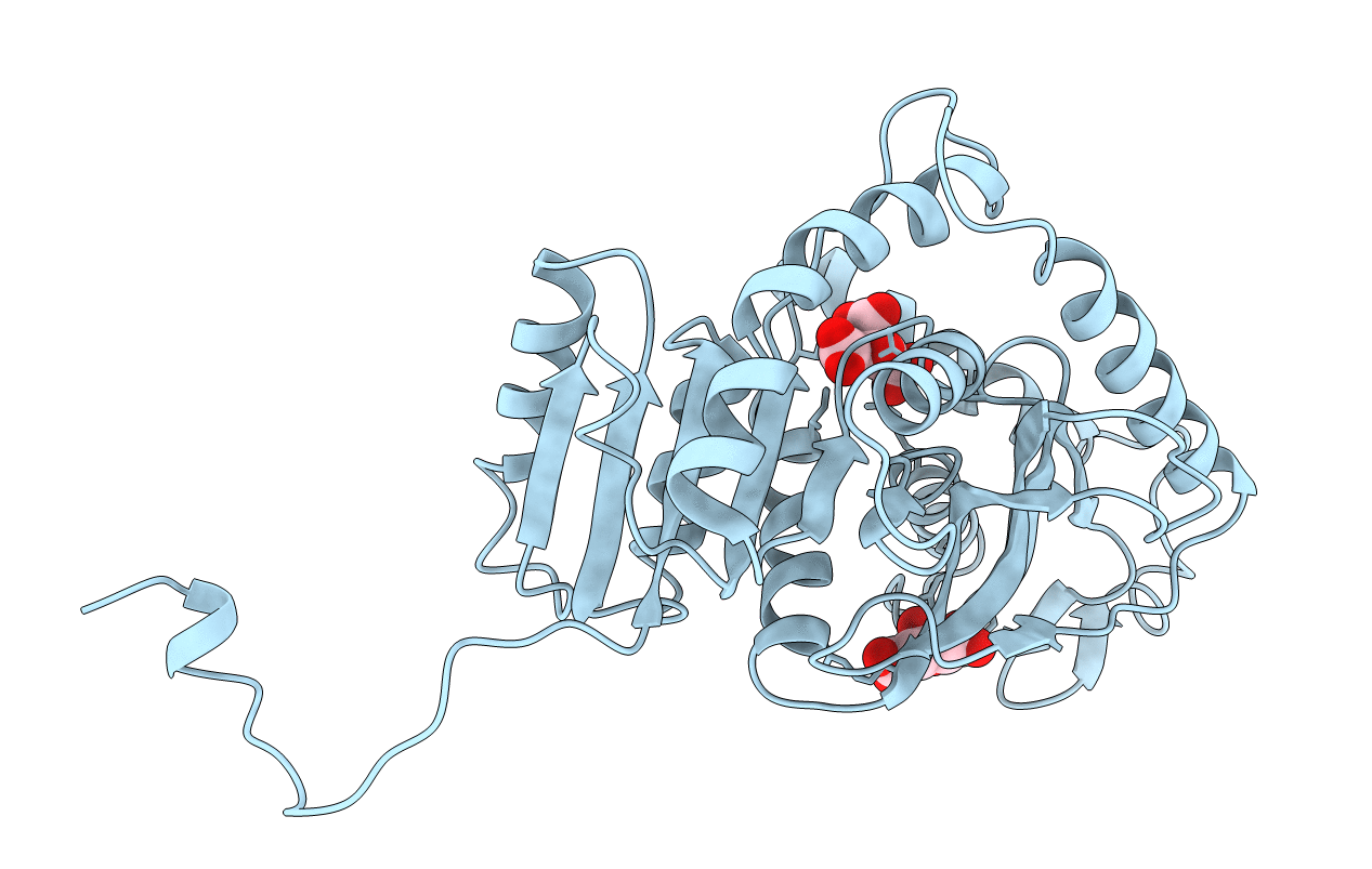
PDB ID:
8LDH
Keywords:
Title:
REFINED CRYSTAL STRUCTURE OF DOGFISH M4 APO-LACTATE DEHYDROGENASE
Biological Source:
Source Organism(s):
Squalus acanthias (Taxon ID: 7797)
Method Details:
Experimental Method:
Resolution:
2.80 Å
R-Value Observed:
0.19
Space Group:
F 4 2 2
