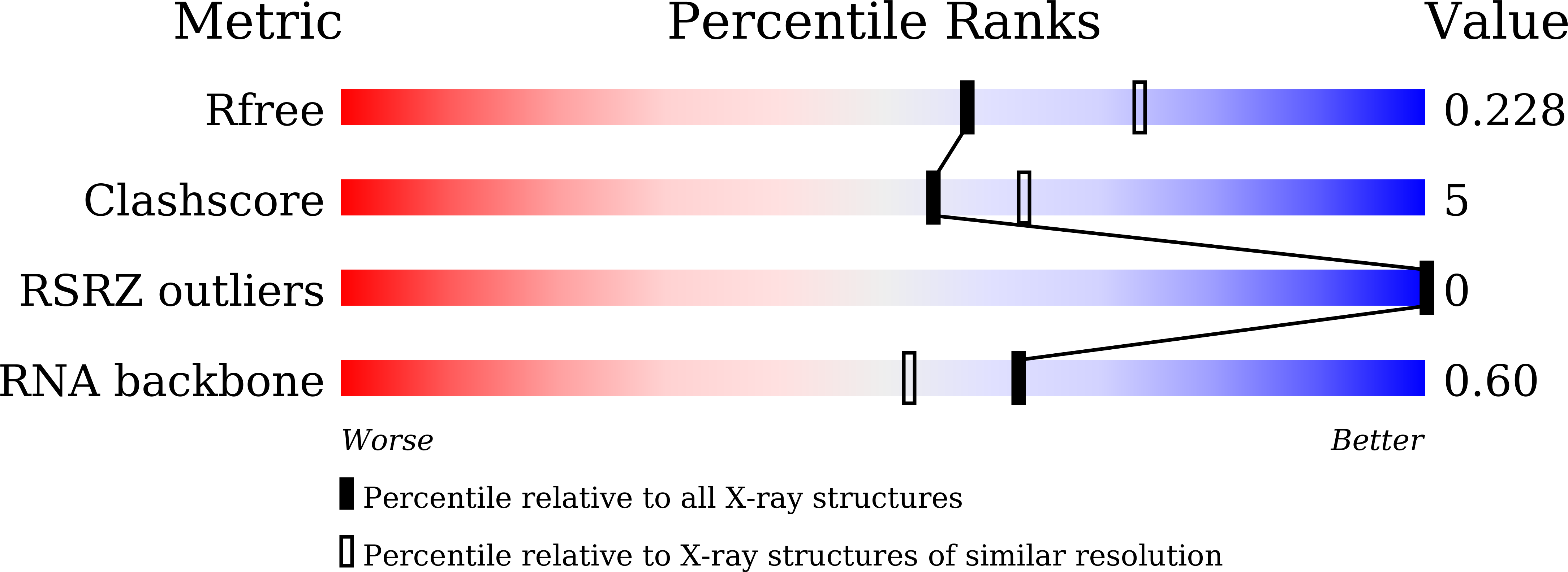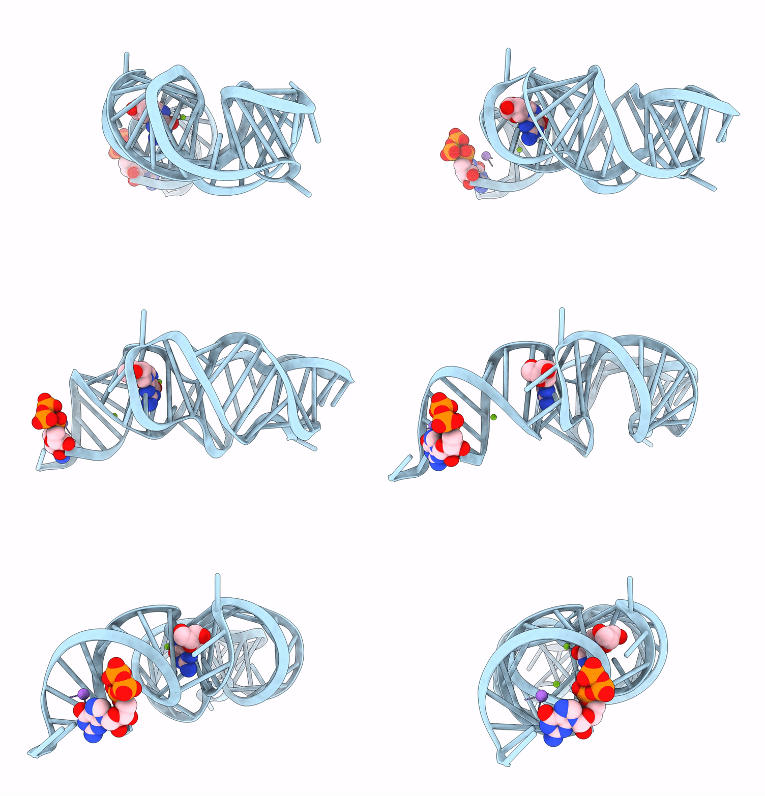
8KEB image
Deposition Date 2023-08-11
Release Date 2025-02-19
Last Version Date 2025-09-10
Entry Detail
PDB ID:
8KEB
Keywords:
Title:
Crystal structure of 2'-dG-III riboswitch with 2'-dG
Biological Source:
Source Organism:
Bacillus sp. (Taxon ID: 1409)
Method Details:
Experimental Method:
Resolution:
2.20 Å
R-Value Free:
0.24
R-Value Work:
0.20
R-Value Observed:
0.20
Space Group:
C 1 2 1
Macromolecular Entities
Polymer Type:polyribonucleotide
Molecule:RNA (72-MER)
Chain IDs:A
Chain Length:72
Number of Molecules:1
Biological Source:Bacillus sp.
Primary Citation
Crystal structures reveal the distinct features of the 2'-dG-III riboswitch in the purine riboswitch family.
Nucleic Acids Res. 53 ? ? (2025)
PMID: 40694847 DOI: 10.1093/nar/gkaf702

Abstact

Purine riboswitches, located in the 5'-untranslated regions of bacterial messenger RNA, regulate gene expression by sensing purines and their derivatives. A class of the guanine-I riboswitch variants was recently reported to be the third class of 2'-deoxyguanosine (2'-dG) riboswitch termed 2'-dG-III riboswitch. Here we present the crystal structures of the 2'-dG-III riboswitch bound with 2'-dG, guanosine, or guanine. Despite similarities in secondary and overall structures to other purine riboswitches, the 2'-dG-III riboswitch exhibits unique features in its loop-loop interaction, three-way junction, and ligand binding. The 2'-dG-III riboswitch exhibits a tuning fork-like structure with a unique six-tiered interaction within the three-way junction. The second, third, and fourth tiers form the ligand-binding pocket, with a consistent binding mode for the guanine moiety of all three ligands. Structural and biochemical analyses reveal detailed interactions between the 2'-dG-III riboswitch and different ligands, providing insights into its regulatory mechanisms in purine metabolism.

Legend

Protein

Chemical

Disease

Primary Citation of related structures