8K2Y image
Deposition Date 2023-07-14
Release Date 2023-11-29
Last Version Date 2023-11-29
Entry Detail
PDB ID:
8K2Y
Keywords:
Title:
Crystal structure of MucD
Biological Source:
Method Details:
Experimental Method:
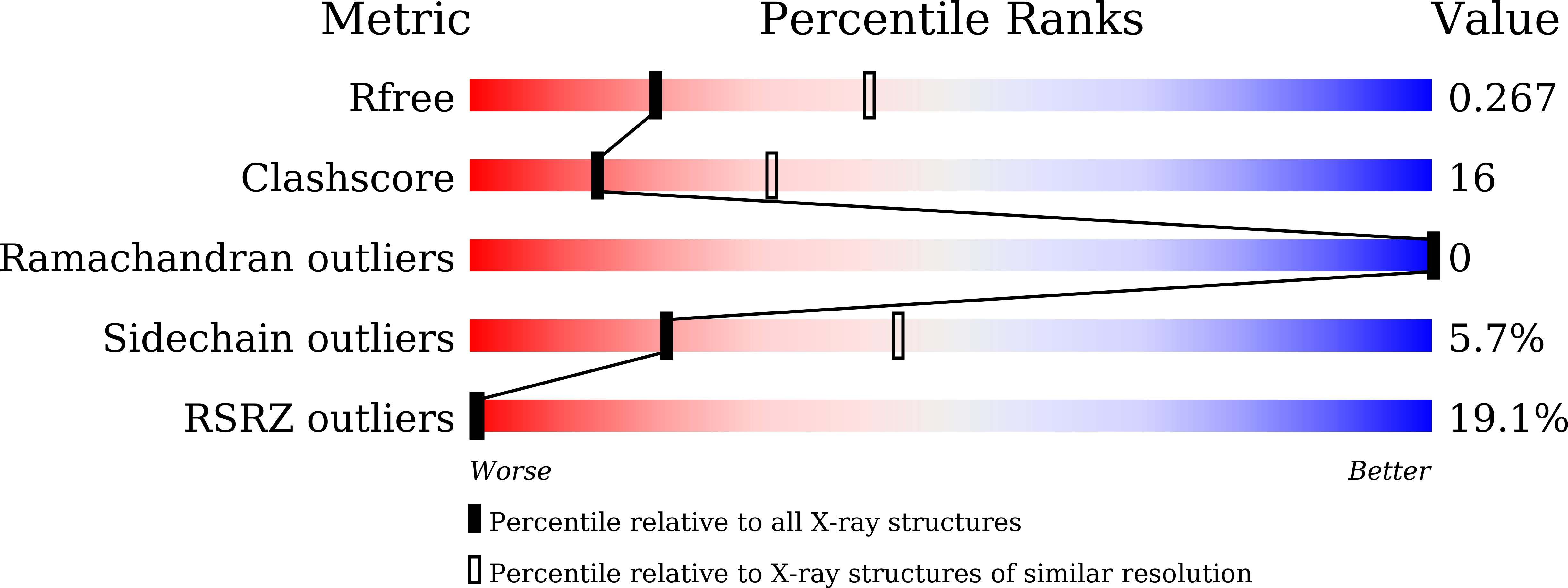
Resolution:
2.70 Å
R-Value Free:
0.26
R-Value Work:
0.20
R-Value Observed:
0.21
Space Group:
C 1 2 1
Macromolecular Entities
Polymer Type:polypeptide(L)
Molecule:serine endoprotease DegP-like protein MucD
Chain IDs:A, B, C
Chain Length:386
Number of Molecules:3
Biological Source:Pseudomonas syringae pv. syringae FF5
Primary Citation
The structure of MucD from Pseudomonas syringae revealed N-terminal loop-mediated trimerization of HtrA-like serine protease.
Biochem.Biophys.Res.Commun. 688 149175 149175 (2023)
PMID: 37976815 DOI: 10.1016/j.bbrc.2023.149175

Abstact

Protein quality control mechanisms are essential for maintaining cellular integrity, and the HtrA family of serine proteases plays a crucial role in handling folding stress in prokaryotic periplasm. Escherichia coli harbors three HtrA members, namely, DegS, DegP, and DegQ, which share a common domain structure. MucD, a putative HtrA family member that resembles DegP, is involved in alginate biosynthesis regulation and the stress response. Pseudomonas syringae causes plant diseases and opportunistic infections in humans. This study presents the high-resolution structure of MucD from Pseudomonas syringae (psMucD), revealing its composition as a typical HtrA family serine protease with protease and PDZ domains. Its findings suggest that psMucD containing one PDZ domain is a trimer in solution, and psMucD trimerization is mediated by its N-terminal loop. Sequence and structural analyses revealed similarities and differences with other HtrA family members. Additionally, this study provides a model of psMucD's catalytic process, comparing it with other members of the HtrA family of serine proteases.

Legend

Protein

Chemical

Disease

Primary Citation of related structures