8K1M image
Deposition Date 2023-07-11
Release Date 2024-07-17
Last Version Date 2025-07-16
Entry Detail
PDB ID:
8K1M
Title:
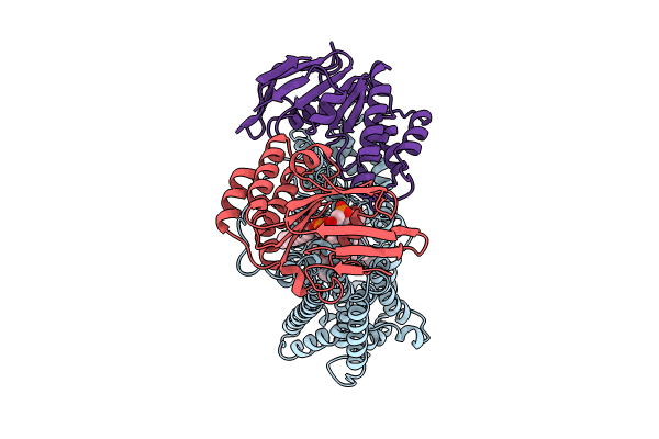
mycobacterial efflux pump, apo state
Biological Source:
Method Details:
Experimental Method:
Resolution:
2.90 Å
Aggregation State:
PARTICLE
Reconstruction Method:
SINGLE PARTICLE
Macromolecular Entities
Structures with similar UniProt ID
Protein Blast
Polymer Type:polypeptide(L)
Molecule:Multidrug efflux system permease protein Rv1217c
Gene (Uniprot):Rv1217c
Chain IDs:A (auth: B)
Chain Length:548
Number of Molecules:1
Biological Source:Mycobacterium tuberculosis (strain ATCC 25618 / H37Rv)
Structures with similar UniProt ID
Protein Blast
Polymer Type:polypeptide(L)
Molecule:Multidrug efflux system ATP-binding protein Rv1218c
Gene (Uniprot):Rv1218c
Chain IDs:B (auth: C), C (auth: D)
Chain Length:289
Number of Molecules:2
Biological Source:Mycobacterium tuberculosis (strain ATCC 25618 / H37Rv)
Primary Citation
Cryo-EM structures of a mycobacterial ABC transporter that mediates rifampicin resistance.
Proc.Natl.Acad.Sci.USA 121 e2403421121 e2403421121 (2024)
PMID: 39226350 DOI: 10.1073/pnas.2403421121

Abstact

Drug-resistant Tuberculosis (TB) is a global public health problem. Resistance to rifampicin, the most effective drug for TB treatment, is a major growing concern. The etiological agent, Mycobacterium tuberculosis (Mtb), has a cluster of ATP-binding cassette (ABC) transporters which are responsible for drug resistance through active export. Here, we describe studies characterizing Mtb Rv1217c-1218c as an ABC transporter that can mediate mycobacterial resistance to rifampicin and have determined the cryo-electron microscopy structures of Rv1217c-1218c. The structures show Rv1217c-1218c has a type V exporter fold. In the absence of ATP, Rv1217c-1218c forms a periplasmic gate by two juxtaposed-membrane helices from each transmembrane domain (TMD), while the nucleotide-binding domains (NBDs) form a partially closed dimer which is held together by four salt-bridges. Adenylyl-imidodiphosphate (AMPPNP) binding induces a structural change where the NBDs become further closed to each other, which downstream translates to a closed conformation for the TMDs. AMPPNP binding results in the collapse of the outer leaflet cavity and the opening of the periplasmic gate, which was proposed to play a role in substrate export. The rifampicin-bound structure shows a hydrophobic and periplasm-facing cavity is involved in rifampicin binding. Phospholipid molecules are observed in all determined structures and form an integral part of the Rv1217c-1218c transporter system. Our results provide a structural basis for a mycobacterial ABC exporter that mediates rifampicin resistance, which can lead to different insights into combating rifampicin resistance.

Legend

Protein

Chemical

Disease

Primary Citation of related structures
Feedback Form
Name
Email
Institute
Feedback