8JYJ image
Entry Detail
PDB ID:
8JYJ
Keywords:
Title:
Crystal structure of engineered HIV-1 Reverse Transcriptase RNase H domain complexed with laccaic acid A
Biological Source:
PDB Version:
Deposition Date:
2023-07-03
Release Date:
2023-08-16
Method Details:
Experimental Method:
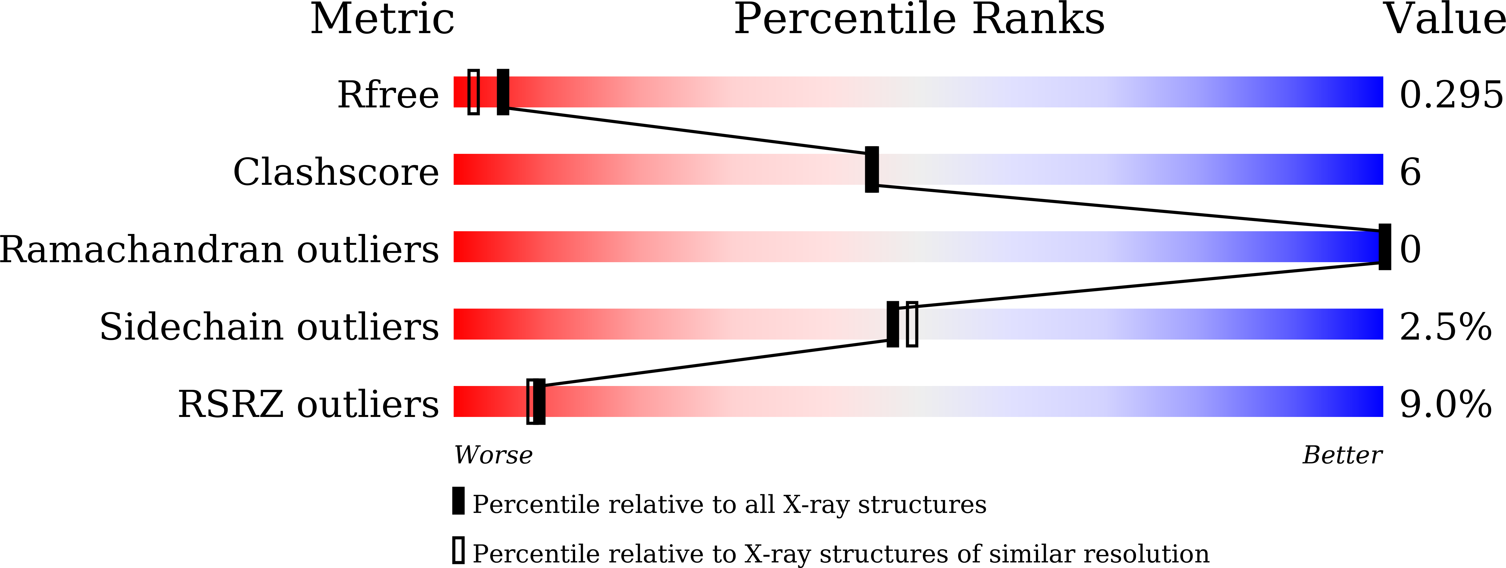
Resolution:
2.01 Å
R-Value Free:
0.29
R-Value Work:
0.24
R-Value Observed:
0.24
Space Group:
P 41 21 2
Macromolecular Entities
Polymer Type:polypeptide(L)
Description:Pol protein,Pol protein,HIV-1 Reverse Transcriptase RNase H active domain
Chain IDs:A
Chain Length:151
Number of Molecules:1
Biological Source:HIV-1 06TG.HT008
Primary Citation
Sticklac-Derived Natural Compounds Inhibiting RNase H Activity of HIV-1 Reverse Transcriptase.
J.Nat.Prod. 86 2487 2495 (2023)
PMID: 37874155 DOI: 10.1021/acs.jnatprod.3c00662

Abstact

The emergence of drug-resistant viruses is a serious concern in current chemotherapy for human immunodeficiency virus type-1 (HIV-1) infectious diseases. Hence, antiviral drugs aiming at targets that are different from those of approved drugs are still required, and the RNase H activity of HIV-1 reverse transcriptase is a suitable target. In this study, a search of a series of natural compounds was performed to identify the RNase H inhibitors. Three compounds were found to block the RNase H enzymatic activity. A laccaic acid skeleton was observed in all three natural compounds. A hydroxy phenyl group is connected to an anthraquinone backbone in the skeleton. An acetamido-ethyl, amino-carboxy-ethyl, and amino-ethyl are bound to the phenyl in laccaic acids A, C, and E, respectively. Laccaic acid C showed a 50% inhibitory concentration at 8.1 μM. Laccaic acid C also showed inhibitory activity in a cell-based viral proliferation assay. Binding structures of these three laccaic acids were determined by X-ray crystallographic analysis using a recombinant protein composed of the HIV-1 RNase H domain. Two divalent metal ions were located at the catalytic center in which one carbonyl and two hydroxy groups on the anthraquinone backbone chelated two metal ions. Molecular dynamics simulations were performed to examine the stabilities of the binding structures. Laccaic acid C showed the strongest binding to the catalytic site. These findings will be helpful for the design of potent inhibitors with modification of laccaic acids to enhance the binding affinity.

Legend

Protein

Chemical

Disease

Primary Citation of related structures