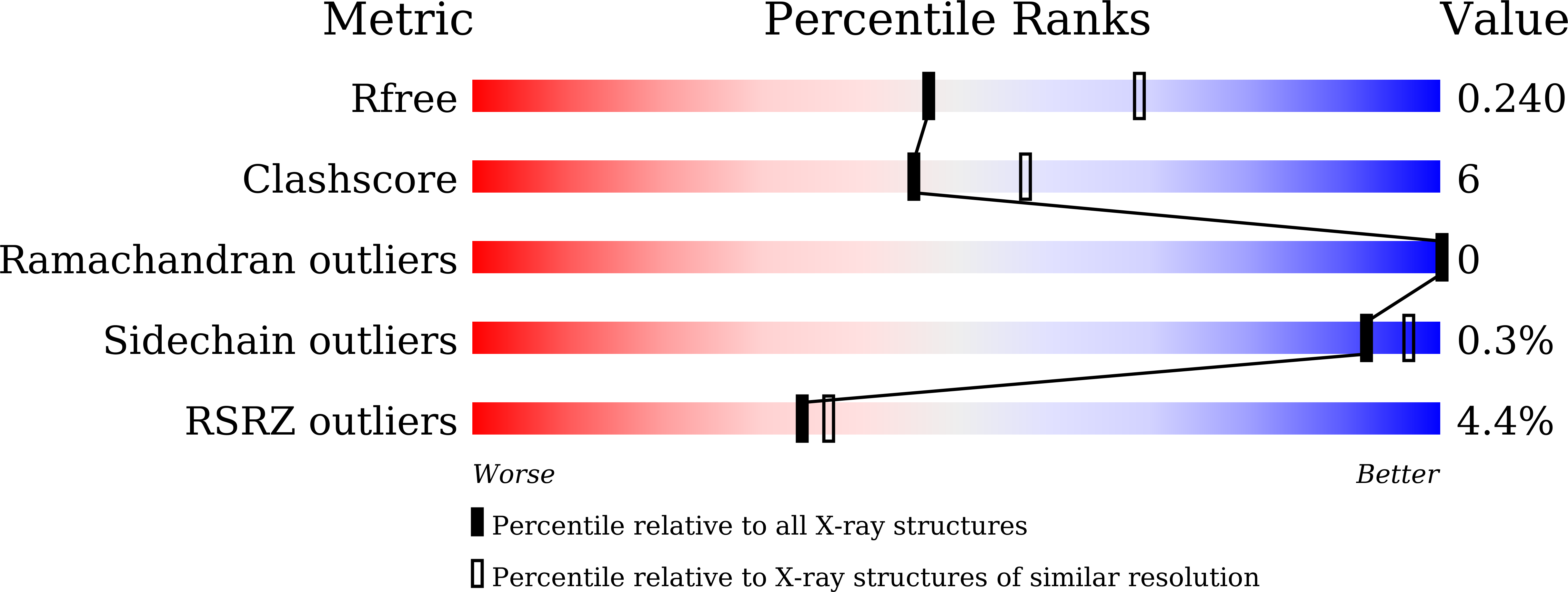
8JWZ image
Entry Detail
PDB ID:
8JWZ
Title:
Crystal structure of A2AR-T4L in complex with AB928
Biological Source:
PDB Version:
Deposition Date:
2023-06-29
Release Date:
2023-08-16
Method Details:
Experimental Method:
Resolution:
2.37 Å
R-Value Free:
0.24
R-Value Work:
0.18
R-Value Observed:
0.19
Space Group:
P 1 21 1
Macromolecular Entities
Polymer Type:polypeptide(L)
Description:Adenosine receptor A2a,Endolysin
Mutations:C54T,C97A
Chain IDs:A
Chain Length:481
Number of Molecules:1
Biological Source:Homo sapiens, Enterobacteria phage T4
Primary Citation
Structural insight into the dual-antagonistic mechanism of AB928 on adenosine A 2 receptors.
Sci China Life Sci 67 986 995 (2024)
PMID: 38319473 DOI: 10.1007/s11427-023-2459-8

Abstact

The adenosine subfamily G protein-coupled receptors A2AR and A2BR have been identified as promising cancer immunotherapy candidates. One of the A2AR/A2BR dual antagonists, AB928, has progressed to a phase II clinical trial to treat rectal cancer. However, the precise mechanism underlying its dual-antagonistic properties remains elusive. Herein, we report crystal structures of the A2AR complexed with AB928 and a selective A2AR antagonist 2-118. The structures revealed a common binding mode on A2AR, wherein the ligands established extensive interactions with residues from the orthosteric and secondary pockets. In contrast, the cAMP assay and A2AR and A2BR molecular dynamics simulations indicated that the ligands adopted distinct binding modes on A2BR. Detailed analysis of their chemical structures suggested that AB928 readily adapted to the A2BR pocket, while 2-118 did not due to intrinsic differences. This disparity potentially accounted for the difference in inhibitory efficacy between A2BR and A2AR. This study serves as a valuable structural template for the future development of selective or dual inhibitors targeting A2AR/A2BR for cancer therapy.

Legend

Protein

Chemical

Disease

Primary Citation of related structures