
Deposition Date
2023-06-27
Release Date
2023-11-29
Last Version Date
2025-07-02
Entry Detail
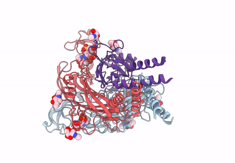
PDB ID:
8JV8
Keywords:
Title:
Cryo-EM structure of the panda P2X7 receptor in complex with PPNDS
Biological Source:
Source Organism(s):
Ailuropoda melanoleuca (Taxon ID: 9646)
Expression System(s):
Method Details:
Experimental Method:
Resolution:
3.34 Å
Aggregation State:
PARTICLE
Reconstruction Method:
SINGLE PARTICLE
