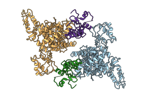
8JRR image
Entry Detail
PDB ID:
8JRR
EMDB ID:
Title:
Structure of E6AP-E6 complex in Det2 state
Biological Source:
PDB Version:
Deposition Date:
2023-06-17
Release Date:
2024-06-05
Method Details:
Experimental Method:
Resolution:
4.35 Å
Aggregation State:
PARTICLE
Reconstruction Method:
SINGLE PARTICLE
Macromolecular Entities
Polymer Type:polypeptide(L)
Description:Ubiquitin-protein ligase E3A
Mutations:T798A/D799A/C843A
Chain IDs:A, C
Chain Length:875
Number of Molecules:2
Biological Source:Homo sapiens
Polymer Type:polypeptide(L)
Description:Protein E6
Mutations:C87S/C104S/C118S
Chain IDs:B, D
Chain Length:135
Number of Molecules:2
Biological Source:Human papillomavirus 16
Ligand Molecules
Primary Citation
Structural insights into the functional mechanism of the ubiquitin ligase E6AP.
Nat Commun 15 3531 3531 (2024)
PMID: 38670961 DOI: 10.1038/s41467-024-47586-w

Abstact

E6AP dysfunction is associated with Angelman syndrome and Autism spectrum disorder. Additionally, the host E6AP is hijacked by the high-risk HPV E6 to aberrantly ubiquitinate the tumor suppressor p53, which is linked with development of multiple types of cancer, including most cervical cancers. Here we show that E6AP and the E6AP/E6 complex exist, respectively, as a monomer and a dimer of the E6AP/E6 protomer. The short α1-helix of E6AP transforms into a longer helical structure when in complex with E6. The extended α1-helices of the dimer intersect symmetrically and contribute to the dimerization. The two protomers sway around the crossed region of the two α1-helices to promote the attachment and detachment of substrates to the catalytic C-lobe of E6AP, thus facilitating ubiquitin transfer. These findings, complemented by mutagenesis analysis, suggest that the α1-helix, through conformational transformations, controls the transition between the inactive monomer and the active dimer of E6AP.

Legend

Protein

Chemical

Disease

Primary Citation of related structures