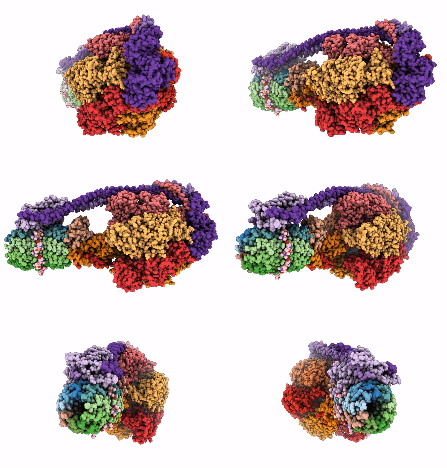
8JR0 image
Entry Detail
PDB ID:
8JR0
EMDB ID:
Title:
Cryo-EM structure of Mycobacterium tuberculosis ATP synthase in complex with TBAJ-587
Biological Source:
PDB Version:
Deposition Date:
2023-06-15
Release Date:
2024-05-01
Method Details:
Experimental Method:
Resolution:
2.80 Å
Aggregation State:
PARTICLE
Reconstruction Method:
SINGLE PARTICLE
Macromolecular Entities
Polymer Type:polypeptide(L)
Description:ATP synthase subunit c
Chain IDs:I (auth: 1), J (auth: 2), K (auth: 3), L (auth: 4), M (auth: 5), N (auth: 6), O (auth: 7), P (auth: 8), Q (auth: 9)
Chain Length:81
Number of Molecules:9
Biological Source:Mycobacterium tuberculosis
Polymer Type:polypeptide(L)
Description:ATP synthase subunit alpha
Chain IDs:A, B, C
Chain Length:549
Number of Molecules:3
Biological Source:Mycobacterium tuberculosis
Polymer Type:polypeptide(L)
Description:ATP synthase subunit beta
Chain IDs:D, E, F
Chain Length:446
Number of Molecules:3
Biological Source:Mycobacterium tuberculosis
Polymer Type:polypeptide(L)
Description:ATP synthase gamma chain
Chain IDs:G
Chain Length:305
Number of Molecules:1
Biological Source:Mycobacterium tuberculosis
Polymer Type:polypeptide(L)
Description:ATP synthase epsilon chain
Chain IDs:H
Chain Length:121
Number of Molecules:1
Biological Source:Mycobacterium tuberculosis
Polymer Type:polypeptide(L)
Description:ATP synthase subunit a
Chain IDs:R (auth: a)
Chain Length:549
Number of Molecules:1
Biological Source:Mycobacterium tuberculosis
Polymer Type:polypeptide(L)
Description:ATP synthase subunit b
Chain IDs:S (auth: b)
Chain Length:549
Number of Molecules:1
Biological Source:Mycobacterium tuberculosis
Polymer Type:polypeptide(L)
Description:Multifunctional fusion protein
Chain IDs:T (auth: d)
Chain Length:446
Number of Molecules:1
Biological Source:Mycobacterium tuberculosis
Primary Citation
Inhibition of M. tuberculosis and human ATP synthase by BDQ and TBAJ-587.
Nature 631 409 414 (2024)
PMID: 38961288 DOI: 10.1038/s41586-024-07605-8

Abstact

Bedaquiline (BDQ), a first-in-class diarylquinoline anti-tuberculosis drug, and its analogue, TBAJ-587, prevent the growth and proliferation of Mycobacterium tuberculosis by inhibiting ATP synthase1,2. However, BDQ also inhibits human ATP synthase3. At present, how these compounds interact with either M. tuberculosis ATP synthase or human ATP synthase is unclear. Here we present cryogenic electron microscopy structures of M. tuberculosis ATP synthase with and without BDQ and TBAJ-587 bound, and human ATP synthase bound to BDQ. The two inhibitors interact with subunit a and the c-ring at the leading site, c-only sites and lagging site in M. tuberculosis ATP synthase, showing that BDQ and TBAJ-587 have similar modes of action. The quinolinyl and dimethylamino units of the compounds make extensive contacts with the protein. The structure of human ATP synthase in complex with BDQ reveals that the BDQ-binding site is similar to that observed for the leading site in M. tuberculosis ATP synthase, and that the quinolinyl unit also interacts extensively with the human enzyme. This study will improve researchers' understanding of the similarities and differences between human ATP synthase and M. tuberculosis ATP synthase in terms of the mode of BDQ binding, and will allow the rational design of novel diarylquinolines as anti-tuberculosis drugs.

Legend

Protein

Chemical

Disease

Primary Citation of related structures