Planned Maintenance: Some services may turn out to be unavailable from 15th January, 2026 to 16th January, 2026. We apologize for the inconvenience!

8JHW image
Deposition Date 2023-05-25
Release Date 2024-05-29
Last Version Date 2025-12-10
Entry Detail
PDB ID:
8JHW
Keywords:
Title:
The first crystal structure of a H-2Kb-restricted decapeptide from Cryptosporidium parvum
Biological Source:
Source Organism(s):
Expression System(s):
Method Details:
Experimental Method:
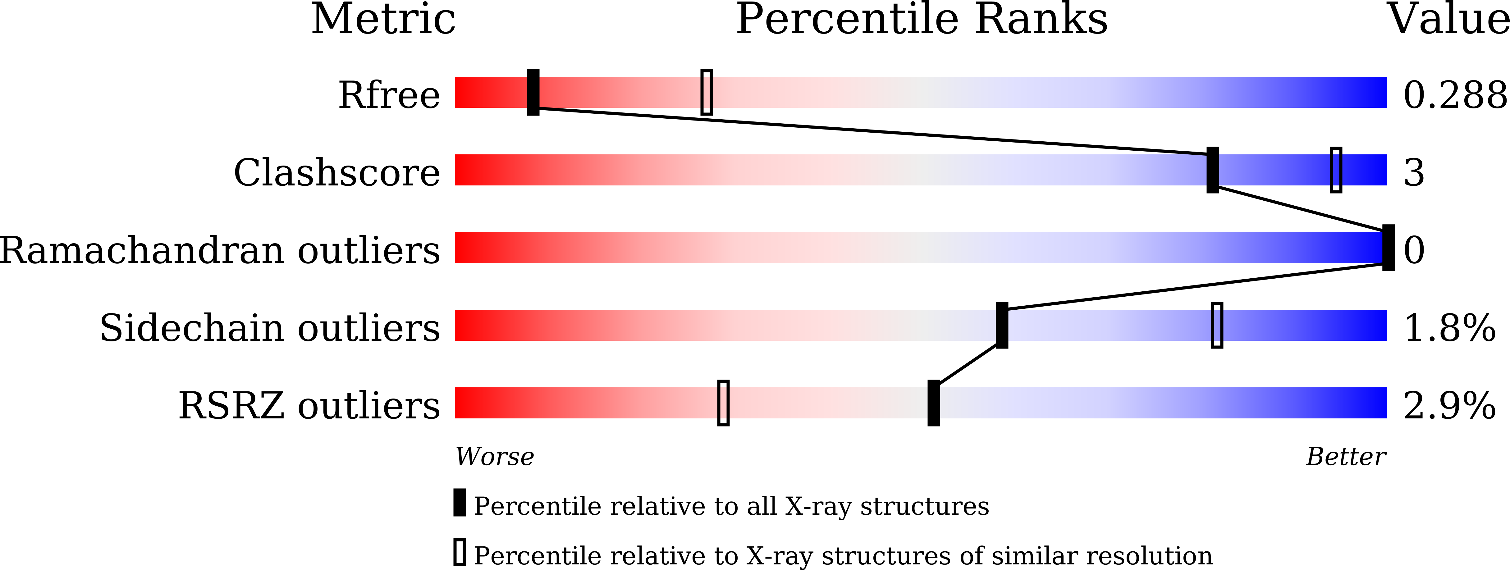
Resolution:
3.12 Å
R-Value Free:
0.28
R-Value Work:
0.24
Space Group:
P 65 2 2
Macromolecular Entities
Structures with similar UniProt ID
Protein Blast
Polymer Type:polypeptide(L)
Molecule:H-2 class I histocompatibility antigen, K-B alpha chain
Gene (Uniprot):H2-K1
Chain IDs:A
Chain Length:275
Number of Molecules:1
Biological Source:Mus musculus
Structures with similar UniProt ID
Protein Blast
Polymer Type:polypeptide(L)
Molecule:Beta-2-microglobulin
Gene (Uniprot):B2m
Chain IDs:B
Chain Length:100
Number of Molecules:1
Biological Source:Mus musculus
Structures with similar UniProt ID
Protein Blast
Polymer Type:polypeptide(L)
Molecule:VAL-THR-PHE-GLU-LYS-SER-TYR-ASN-THR-VAL
Chain IDs:C
Chain Length:10
Number of Molecules:1
Biological Source:Cryptosporidium parvum
Ligand Molecules
Primary Citation
Structural analyses of Cryptosporidium parvum epitopes reveal a novel scheme of decapeptide binding to H-2K b.
J.Struct.Biol. 217 108168 108168 (2025)
PMID: 39809366 DOI: 10.1016/j.jsb.2025.108168

Abstact

Cryptosporidium has gained much attention as a major cause of diarrhea worldwide. Here, we present the first structure of H-2Kb complexed with a decapeptide from Cryptosporidium parvum Gp40/15 protein (Gp40/15-VTF10). In contrast to all published structures, the aromatic residue P3-Phe of Gp40/15-VTF10 is anchored in pocket C rather than the canonical Y/F at P5 or P6 reported for octapeptides and nonapeptides. The results of in vitro refolding assays and circular dichroism experiments showed that the side chains of P3 and P5 play key roles in Gp40/15-VTF10 peptide binding. However, functional analysis of decapeptide epitopes revealed that the Gp40/15-VTF10 peptide did not elicit a strong CD8+T immune response, whereas the decapeptide epitope MEDLE2-INF10 induced a significant CD8+ T-cell response in peptide-immunized C57BL/6 mice. Using a model structure of H-2Kb-INF10 complex, we found that the antigenic decapeptide INF10 exhibits a completely different conformation, with the aromatic anchors P3F and P7F docked into the D and C pockets, respectively, while similar peptide conformation and hydrogen bond interactions between the peptide and major histocompatibility complex were found in the resolved H-2Kb-SVF9 complex. As the H-2Kb molecule predominantly prefers octapeptides with a strong anchor of P5 Y/F (or P6 Y/F for nonapeptides) binding to the C pocket, we propose that P7 Y/F in the C pocket may represent a novel binding mode for decapeptides. The results should increase the accuracy of T-cell epitope prediction and support the development of T-cell epitope vaccines against cryptosporidiosis.

Legend

Protein

Chemical

Disease

Primary Citation of related structures
Feedback Form
Name
Email
Institute
Feedback