8JG2 image
Deposition Date 2023-05-19
Release Date 2024-05-08
Last Version Date 2025-11-19
Entry Detail
PDB ID:
8JG2
Title:
Crystal structure of a biosynthetic thiolase from Megasphaera hexanoica soaked with hexanoyl-CoA
Biological Source:
Source Organism:
Method Details:
Experimental Method:
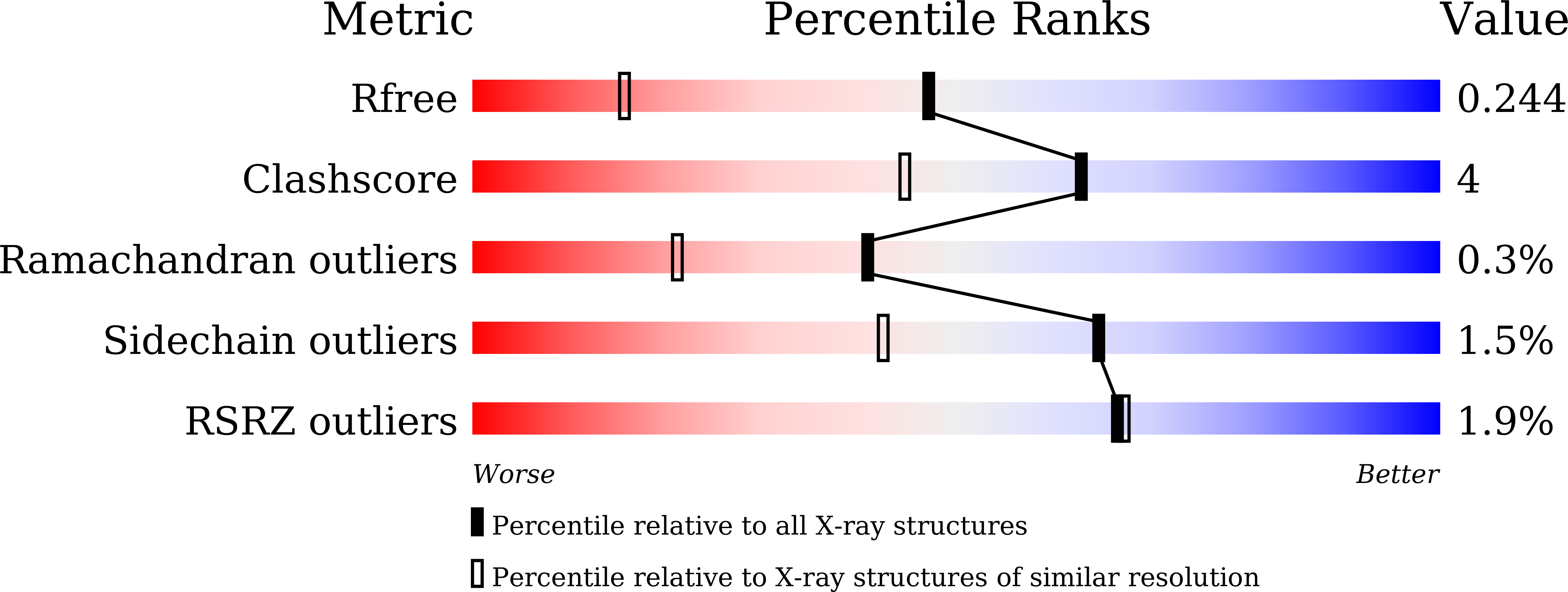
Resolution:
1.64 Å
R-Value Free:
0.23
R-Value Work:
0.19
R-Value Observed:
0.20
Space Group:
P 2 21 21
Macromolecular Entities
Structures with similar UniProt ID
Protein Blast
Polymer Type:polypeptide(L)
Molecule:Acetyl-CoA C-acetyltransferase
Gene (Uniprot):HF872_02440
Mutagens:C88S
Chain IDs:A, B
Chain Length:434
Number of Molecules:2
Biological Source:Megasphaera hexanoica
Primary Citation
Molecular Chain Elongation Mechanism for n-Caproate Biosynthesis by Megasphaera Hexanoica.
Adv Sci ? e06069 e06069 (2025)
PMID: 40932660 DOI: 10.1002/advs.202506069

Abstact

The microbial production of medium-chain carboxylates has attracted considerable interest owing to their potential applications in biofuels and specialty chemicals; however, the underlying biosynthetic mechanisms remain incompletely understood. The present study evaluates the medium-chain carboxylate-producing microbe Megaspahera hexanoica using genomic analysis, transcriptome analysis, and metabolic engineering. Additionally, the n-caproate synthesis pathway of M. hexanoica is characterized with fructose as an electron donor, and the substrate specificity of the respective proteins is evaluated by constructing an n-caproate biosynthetic pathway in Escherichia coli. Among all r-BOX or RBO genes, thl_1583, which encodes β-ketothiolase (MhTHL), is identified as the most critical enzyme for the carbon chain elongation mechanism in M. hexanoica. Therefore, MhTHL is compared with other well-studied β-ketothiolases (CkTHL from Clostridium kluyveri, ReBktB from Ralstonia eutropha (Cupriavidus necator), EcAtoB from E. coli, and CaTHL from C. acetobutylicum). MhTHL is found to exhibit the highest n-caproate production, as evidenced by the protein crystal structure of MhTHL. Structural comparisons with other thiolases show that MhTHL has a larger substrate-binding pocket than ReBktB. Thiolase mutants generated by site-directed mutagenesis reveal that two residues (Leu87 and Val351) are essential for determining the size of the substrate-binding pocket.

Legend

Protein

Chemical

Disease

Primary Citation of related structures
Feedback Form
Name
Email
Institute
Feedback