8JFM image
Entry Detail
PDB ID:
8JFM
Title:
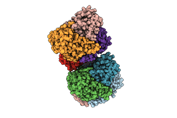
Crystal structure of enoyl-ACP reductase FabI in complex with NADH from Helicobacter pylori
Biological Source:
Source Organism:
Host Organism:
PDB Version:
Deposition Date:
2023-05-18
Release Date:
2023-11-15
Method Details:
Experimental Method:
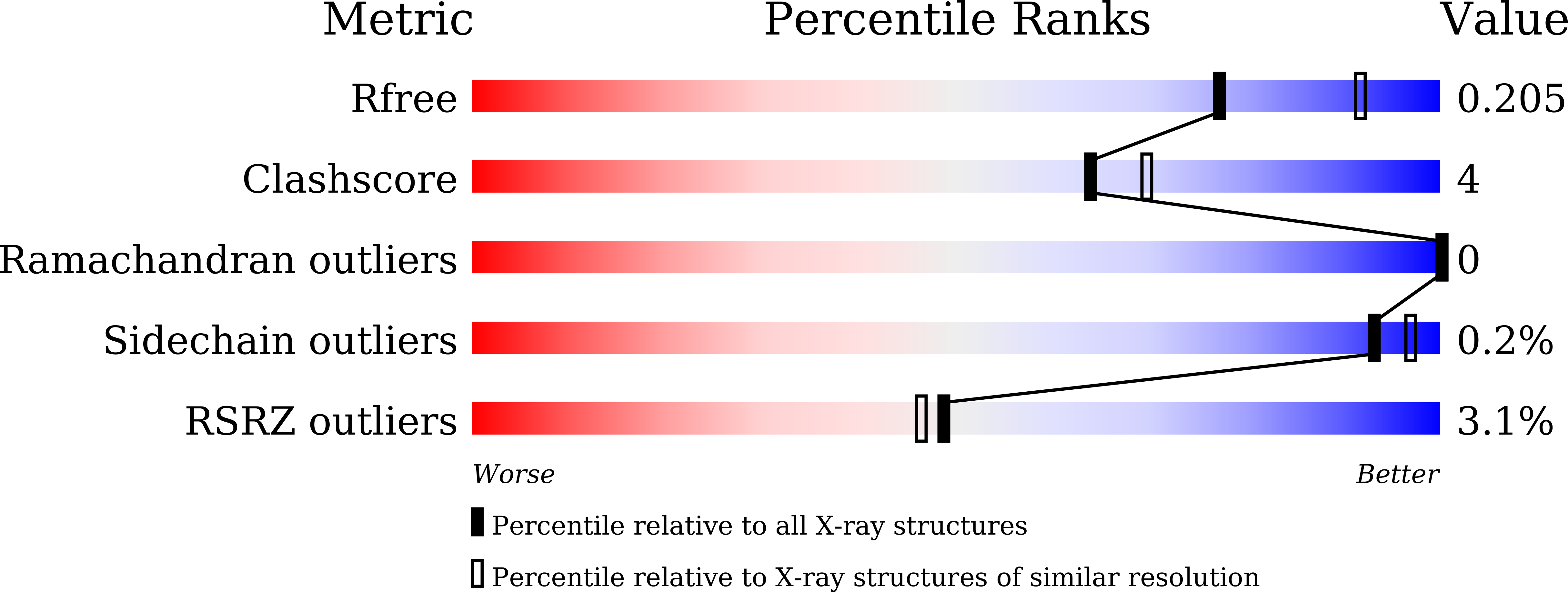
Resolution:
2.21 Å
R-Value Free:
0.20
R-Value Work:
0.15
R-Value Observed:
0.15
Space Group:
P 1 21 1
Macromolecular Entities
Polymer Type:polypeptide(L)
Description:Enoyl-[acyl-carrier-protein] reductase [NADH]
Chain IDs:A, B, C, D, E, F, G, H
Chain Length:273
Number of Molecules:8
Biological Source:Helicobacter pylori
Ligand Molecules
Primary Citation
The Molecular Basis of Catalysis by SDR Family Members Ketoacyl-ACP Reductase FabG and Enoyl-ACP Reductase FabI in Type-II Fatty Acid Biosynthesis.
Angew.Chem.Int.Ed.Engl. 62 e202313109 e202313109 (2023)
PMID: 37779101 DOI: 10.1002/anie.202313109

Abstact

The short-chain dehydrogenase/reductase (SDR) superfamily members acyl-ACP reductases FabG and FabI are indispensable core enzymatic modules and catalytic orientation controllers in type-II fatty acid biosynthesis. Herein, we report their distinct substrate allosteric recognition and enantioselective reduction mechanisms. FabG achieves allosteric regulation of ACP and NADPH through ACP binding across two adjacent FabG monomers, while FabI follows an irreversible compulsory order of substrate binding in that NADH binding must precede that of ACP on a discrete FabI monomer. Moreover, FabG and FabI utilize a backdoor residue Phe187 or a "rheostat" α8 helix for acyl chain length selection, and their corresponding triad residues Ser142 or Tyr145 recognize the keto- or enoyl-acyl substrates, respectively, facilitating initiation of nucleophilic attack by NAD(P)H. The other two triad residues (Tyr and Lys) mediate subsequent proton transfer and (R)-3-hydroxyacyl- or saturated acyl-ACP production.

Legend

Protein

Chemical

Disease

Primary Citation of related structures