
Deposition Date
2023-05-15
Release Date
2023-10-25
Last Version Date
2024-01-17
Entry Detail
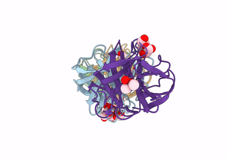
PDB ID:
8JEA
Keywords:
Title:
Crystal structure of CGL1 from Crassostrea gigas, mannotriose-bound form (CGL1/Man(alpha)1-2Man(alpha)1-2Man)
Biological Source:
Source Organism(s):
Crassostrea gigas (Taxon ID: 29159)
Method Details:
Experimental Method:
Resolution:
0.97 Å
R-Value Free:
0.18
R-Value Work:
0.17
Space Group:
P 1 21 1
