8JD3 image
Deposition Date 2023-05-12
Release Date 2023-06-21
Last Version Date 2024-11-06
Entry Detail
PDB ID:
8JD3
Title:
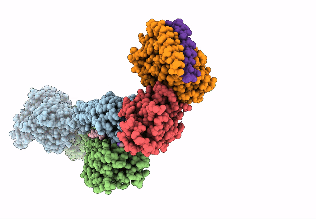
Cryo-EM structure of Gi1-bound mGlu2-mGlu3 heterodimer
Biological Source:
Source Organism:
Homo sapiens (Taxon ID: 9606)
Method Details:
Experimental Method:
Resolution:
3.30 Å
Aggregation State:
PARTICLE
Reconstruction Method:
SINGLE PARTICLE
Macromolecular Entities
Structures with similar UniProt ID
Protein Blast
Polymer Type:polypeptide(L)
Molecule:Metabotropic glutamate receptor 2
Gene (Uniprot):GRM2
Chain IDs:A (auth: 2)
Chain Length:870
Number of Molecules:1
Biological Source:Homo sapiens
Structures with similar UniProt ID
Protein Blast
Polymer Type:polypeptide(L)
Molecule:Metabotropic glutamate receptor 3
Gene (Uniprot):GRM3
Chain IDs:B (auth: 3)
Chain Length:894
Number of Molecules:1
Biological Source:Homo sapiens
Structures with similar UniProt ID
Protein Blast
Polymer Type:polypeptide(L)
Molecule:Guanine nucleotide-binding protein G(i) subunit alpha-1
Gene (Uniprot):GNAI1
Chain IDs:C (auth: A)
Chain Length:354
Number of Molecules:1
Biological Source:Homo sapiens
Structures with similar UniProt ID
Protein Blast
Polymer Type:polypeptide(L)
Molecule:Guanine nucleotide-binding protein G(I)/G(S)/G(T) subunit beta-1
Gene (Uniprot):GNB1
Chain IDs:D (auth: B)
Chain Length:351
Number of Molecules:1
Biological Source:Homo sapiens
Structures with similar UniProt ID
Protein Blast
Polymer Type:polypeptide(L)
Molecule:Guanine nucleotide-binding protein G(I)/G(S)/G(O) subunit gamma-2
Gene (Uniprot):GNG2
Chain IDs:E (auth: C)
Chain Length:71
Number of Molecules:1
Biological Source:Homo sapiens
Primary Citation
Structural insights into dimerization and activation of the mGlu2-mGlu3 and mGlu2-mGlu4 heterodimers.
Cell Res. 33 762 774 (2023)
PMID: 37286794 DOI: 10.1038/s41422-023-00830-2

Abstact

Heterodimerization of the metabotropic glutamate receptors (mGlus) has shown importance in the functional modulation of the receptors and offers potential drug targets for treating central nervous system diseases. However, due to a lack of molecular details of the mGlu heterodimers, understanding of the mechanisms underlying mGlu heterodimerization and activation is limited. Here we report twelve cryo-electron microscopy (cryo-EM) structures of the mGlu2-mGlu3 and mGlu2-mGlu4 heterodimers in different conformational states, including inactive, intermediate inactive, intermediate active and fully active conformations. These structures provide a full picture of conformational rearrangement of mGlu2-mGlu3 upon activation. The Venus flytrap domains undergo a sequential conformational change, while the transmembrane domains exhibit a substantial rearrangement from an inactive, symmetric dimer with diverse dimerization patterns to an active, asymmetric dimer in a conserved dimerization mode. Combined with functional data, these structures reveal that stability of the inactive conformations of the subunits and the subunit-G protein interaction pattern are determinants of asymmetric signal transduction of the heterodimers. Furthermore, a novel binding site for two mGlu4 positive allosteric modulators was observed in the asymmetric dimer interfaces of the mGlu2-mGlu4 heterodimer and mGlu4 homodimer, and may serve as a drug recognition site. These findings greatly extend our knowledge about signal transduction of the mGlus.

Legend

Protein

Chemical

Disease

Primary Citation of related structures