Deposition Date
2023-05-08
Release Date
2023-11-15
Last Version Date
2023-11-15
Entry Detail
PDB ID:
8JBD
Keywords:
Title:
Crystal structure of Adenylosuccinate lyase from Thermus thermophilus HB8, TtPurB
Biological Source:
Source Organism(s):
Thermus thermophilus HB8 (Taxon ID: 300852)
Expression System(s):
Method Details:
Experimental Method:
Resolution:
2.38 Å
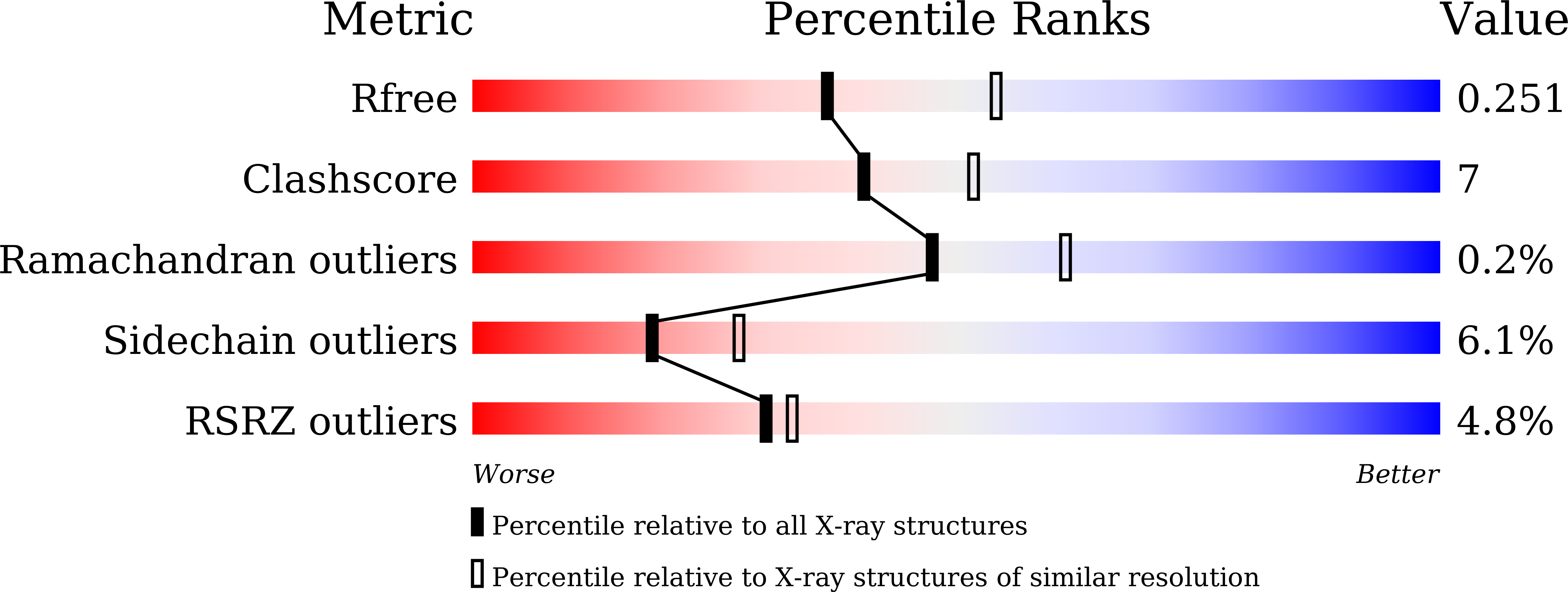
R-Value Free:
0.24
R-Value Work:
0.20
Space Group:
P 65 2 2