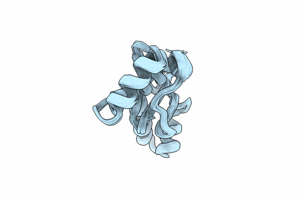
8JB9 image
Entry Detail
PDB ID:
8JB9
Keywords:
Title:
Solution structure of Anti-CRISPR protein AcrIIC5
Biological Source:
Source Organism:
PDB Version:
Deposition Date:
2023-05-08
Release Date:
2024-05-08
Method Details:
Experimental Method:
Conformers Calculated:
1800
Conformers Submitted:
20
Selection Criteria:
structures with the lowest energy
Macromolecular Entities
Polymer Type:polypeptide(L)
Description:Type II-C anti-CRISPR protein, AcrIIC5
Chain IDs:A
Chain Length:130
Number of Molecules:1
Biological Source:Simonsiella muelleri
Ligand Molecules
Primary Citation
Structural variants of AcrIIC5 inhibit Cas9 via divergent binding interfaces.
Structure 33 517 527.e5 (2025)
PMID: 39826547 DOI: 10.1016/j.str.2024.12.014

Abstact

CRISPR-Cas is a bacterial defense system that employs RNA-guided endonucleases to destroy invading foreign nucleic acids. Bacteriophages produce anti-CRISPR (Acr) proteins to evade CRISPR-Cas defense during the infection. AcrIIC5, a type II-C Cas9 inhibitor, exhibits unusual variations in the local backbone fold between its orthologs. Here we investigated how the folding variations affect the inhibition of target Cas9 using AcrIIC5 orthologs. Structural comparison of free AcrIIC5Smu and AcrIIC5Nch confirmed that the folding variation correlated with characteristic indels in the helical region. Mutagenesis and biochemical assays combined with AlphaFold2 predictions identified key residues of AcrIIC5 orthologs important for Cas9 inhibition. Remarkably, AcrIIC5 orthologs employed divergent binding interfaces via folding variations to inhibit the Cas9 targets. Our study suggests that Acr proteins have evolved structural variants to diversify key interfaces for target Cas9, which could be beneficial for the adaptation of phages to evasive mutations on the Cas9 surface.

Legend

Protein

Chemical

Disease

Primary Citation of related structures