
Deposition Date
2023-05-05
Release Date
2023-12-27
Last Version Date
2024-10-23
Entry Detail
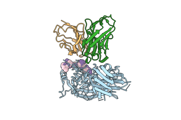
PDB ID:
8JAF
Keywords:
Title:
Structure of Muscarinic receptor (M2R) in complex with beta-arrestin1 (Local Refine, non-cross linked)
Biological Source:
Source Organism(s):
Bos taurus (Taxon ID: 9913)
Mus musculus (Taxon ID: 10090)
Homo sapiens (Taxon ID: 9606)
Mus musculus (Taxon ID: 10090)
Homo sapiens (Taxon ID: 9606)
Expression System(s):
Method Details:
Experimental Method:
Resolution:
3.10 Å
Aggregation State:
PARTICLE
Reconstruction Method:
SINGLE PARTICLE
