8J8Z image
Deposition Date 2023-05-02
Release Date 2023-12-27
Last Version Date 2024-11-06
Entry Detail
PDB ID:
8J8Z
Title:
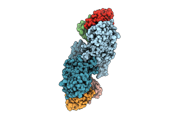
Structure of beta-arrestin1 in complex with D6Rpp
Biological Source:
Source Organism:
Rattus norvegicus (Taxon ID: 10116)
Mus musculus (Taxon ID: 10090)
Homo sapiens (Taxon ID: 9606)
Host Organism:
Method Details:
Experimental Method:
Resolution:
3.40 Å
Aggregation State:
PARTICLE
Reconstruction Method:
SINGLE PARTICLE
Macromolecular Entities
Structures with similar UniProt ID
Protein Blast
Polymer Type:polypeptide(L)
Molecule:Beta-arrestin-1
Gene (Uniprot):Arrb1
Chain IDs:A, E (auth: B)
Chain Length:418
Number of Molecules:2
Biological Source:Rattus norvegicus
Polymer Type:polypeptide(L)
Molecule:Fab30 Heavy Chain
Chain IDs:B (auth: H), F (auth: I)
Chain Length:237
Number of Molecules:2
Biological Source:Mus musculus
Polymer Type:polypeptide(L)
Molecule:Fab30 Light Chain
Chain IDs:C (auth: L), G (auth: M)
Chain Length:215
Number of Molecules:2
Biological Source:Mus musculus
Structures with similar UniProt ID
Protein Blast
Polymer Type:polypeptide(L)
Molecule:Atypical chemokine receptor 2
Gene (Uniprot):ACKR2
Chain IDs:D (auth: V), H (auth: U)
Chain Length:18
Number of Molecules:2
Biological Source:Homo sapiens
Modified Residue
Compound ID Chain ID Parent Comp ID Details 2D Image
SEP D SER modified residue
Ligand Molecules
Primary Citation
Molecular insights into atypical modes of beta-arrestin interaction with seven transmembrane receptors.
Science 383 101 108 (2024)
PMID: 38175886 DOI: 10.1126/science.adj3347

Abstact

β-arrestins (βarrs) are multifunctional proteins involved in signaling and regulation of seven transmembrane receptors (7TMRs), and their interaction is driven primarily by agonist-induced receptor activation and phosphorylation. Here, we present seven cryo-electron microscopy structures of βarrs either in the basal state, activated by the muscarinic receptor subtype 2 (M2R) through its third intracellular loop, or activated by the βarr-biased decoy D6 receptor (D6R). Combined with biochemical, cellular, and biophysical experiments, these structural snapshots allow the visualization of atypical engagement of βarrs with 7TMRs and also reveal a structural transition in the carboxyl terminus of βarr2 from a β strand to an α helix upon activation by D6R. Our study provides previously unanticipated molecular insights into the structural and functional diversity encoded in 7TMR-βarr complexes with direct implications for exploring novel therapeutic avenues.

Legend

Protein

Chemical

Disease

Primary Citation of related structures