8ITS image
Deposition Date 2023-03-22
Release Date 2024-03-27
Last Version Date 2025-04-09
Entry Detail
PDB ID:
8ITS
Keywords:
Title:
Crystal structure of DUF-3268 k-junction
Biological Source:
Source Organism:
bacterium (Taxon ID: 1869227)
Method Details:
Experimental Method:
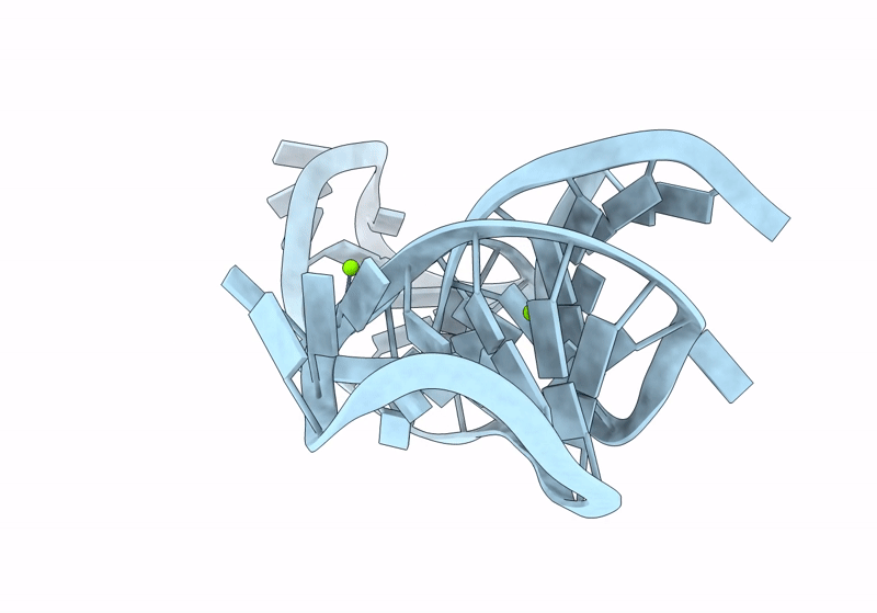
Resolution:
1.94 Å
R-Value Free:
0.22
R-Value Work:
0.20
R-Value Observed:
0.20
Space Group:
C 1 2 1
Macromolecular Entities
Polymer Type:polyribonucleotide
Molecule:RNA (46-MER)
Chain IDs:A
Chain Length:46
Number of Molecules:1
Biological Source:bacterium
Primary Citation
Structure and ion-dependent folding of k-junctions.
Rna 29 1411 1422 (2023)
PMID: 37311599 DOI: 10.1261/rna.079678.123

Abstact

k-Junctions are elaborated forms of kink turns with an additional helix on the nonbulged strand, thus forming a three-way helical junction. Two were originally identified in the structures of Arabidopsis and Escherichia coli thiamine pyrophosphate (TPP) riboswitches, and another called DUF-3268 was tentatively identified from sequence information. In this work we show that the Arabidopsis and E. coli riboswitch k-junctions fold in response to the addition of magnesium or sodium ions, and that atomic mutations that should disrupt key hydrogen bonding interactions greatly impair folding. Using X-ray crystallography, we have determined the structure of the DUF-3268 RNA and thus confirmed that it is a k-junction. It also folds upon the addition of metal ions, though requiring a 40-fold lower concentration of either divalent or monovalent ions. The key difference between the DUF-3268 and riboswitch k-junctions is the lack of nucleotides inserted between G1b and A2b in the former. We show that this insertion is primarily responsible for the difference in folding properties. Finally, we show that the DUF-3268 can functionally substitute for the k-junction in the E. coli TPP riboswitch such that the chimera can bind the TPP ligand, although less avidly.

Legend

Protein

Chemical

Disease

Primary Citation of related structures