8ITL image
Deposition Date 2023-03-22
Release Date 2023-10-18
Last Version Date 2024-11-13
Entry Detail
PDB ID:
8ITL
Title:
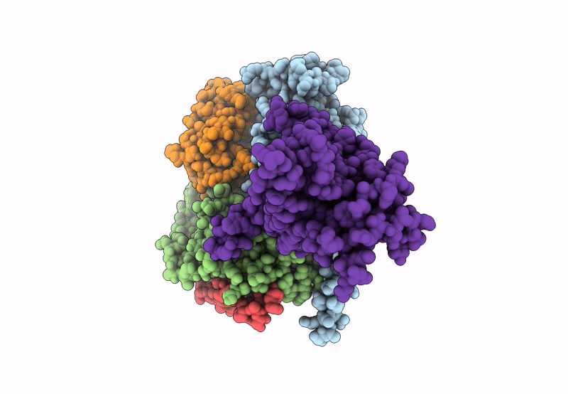
Cryo-EM structure of GIPR splice variant 1 (SV1) in complex with Gs protein
Biological Source:
Source Organism:
Homo sapiens (Taxon ID: 9606)
Bos taurus (Taxon ID: 9913)
Rattus norvegicus (Taxon ID: 10116)
synthetic construct (Taxon ID: 32630)
Host Organism:
Method Details:
Experimental Method:
Resolution:
3.23 Å
Aggregation State:
PARTICLE
Reconstruction Method:
SINGLE PARTICLE
Macromolecular Entities
Polymer Type:polypeptide(L)
Molecule:Guanine nucleotide-binding protein G(s) subunit alpha isoforms short
Gene (Uniprot):GNAS
Mutations:S54N,G226A,E268A,N271K,K274D,R280K,T284D,I285T
Chain IDs:B (auth: A)
Chain Length:394
Number of Molecules:1
Biological Source:Bos taurus
Polymer Type:polypeptide(L)
Molecule:Guanine nucleotide-binding protein G(I)/G(S)/G(T) subunit beta-1
Gene (Uniprot):Gnb1
Chain IDs:C (auth: B)
Chain Length:371
Number of Molecules:1
Biological Source:Rattus norvegicus
Polymer Type:polypeptide(L)
Molecule:Guanine nucleotide-binding protein G(I)/G(S)/G(O) subunit gamma-2
Gene (Uniprot):GNG2
Chain IDs:D (auth: G)
Chain Length:71
Number of Molecules:1
Biological Source:Bos taurus
Polymer Type:polypeptide(L)
Molecule:Nanobody-35
Chain IDs:E (auth: N)
Chain Length:128
Number of Molecules:1
Biological Source:synthetic construct
Polymer Type:polypeptide(L)
Molecule:Gastric inhibitory polypeptide receptor
Gene (Uniprot):GIPR
Mutations:T309F
Chain IDs:A (auth: R)
Chain Length:364
Number of Molecules:1
Biological Source:Homo sapiens
Primary Citation
Molecular basis of signal transduction mediated by the human GIPR splice variants.
Proc.Natl.Acad.Sci.USA 120 e2306145120 e2306145120 (2023)
PMID: 37792509 DOI: 10.1073/pnas.2306145120

Abstact

Glucose-dependent insulinotropic polypeptide receptor (GIPR) is a potential drug target for metabolic disorders. It works with glucagon-like peptide-1 receptor and glucagon receptor in humans to maintain glucose homeostasis. Unlike the other two receptors, GIPR has at least 13 reported splice variants (SVs), more than half of which have sequence variations at either C or N terminus. To explore their roles in endogenous peptide-mediated GIPR signaling, we determined the cryoelectron microscopy (cryo-EM) structures of the two N terminus-altered SVs (referred as GIPR-202 and GIPR-209 in the Ensembl database, SV1 and SV2 here, respectively) and investigated the outcome of coexpressing each of them in question with GIPR in HEK293T cells with respect to ligand binding, receptor expression, cAMP (adenosine 3,5-cyclic monophosphate) accumulation, β-arrestin recruitment, and cell surface localization. It was found that while both N terminus-altered SVs of GIPR neither bound to the hormone nor elicited signal transduction per se, they suppressed ligand binding and cAMP accumulation of GIPR. Meanwhile, SV1 reduced GIPR-mediated β-arrestin 2 responses. The cryo-EM structures of SV1 and SV2 showed that they reorganized the extracellular halves of transmembrane helices 1, 6, and 7 and extracellular loops 2 and 3 to adopt a ligand-binding pocket-occupied conformation, thereby losing binding ability to the peptide. The results suggest a form of signal bias that is constitutive and ligand-independent, thus expanding our knowledge of biased signaling beyond pharmacological manipulation (i.e., ligand specific) as well as constitutive and ligand-independent (e.g., SV1 of the growth hormone-releasing hormone receptor).

Legend

Protein

Chemical

Disease

Primary Citation of related structures