8IM5 image
Deposition Date 2023-03-06
Release Date 2023-08-30
Last Version Date 2024-05-15
Entry Detail
PDB ID:
8IM5
Keywords:
Title:
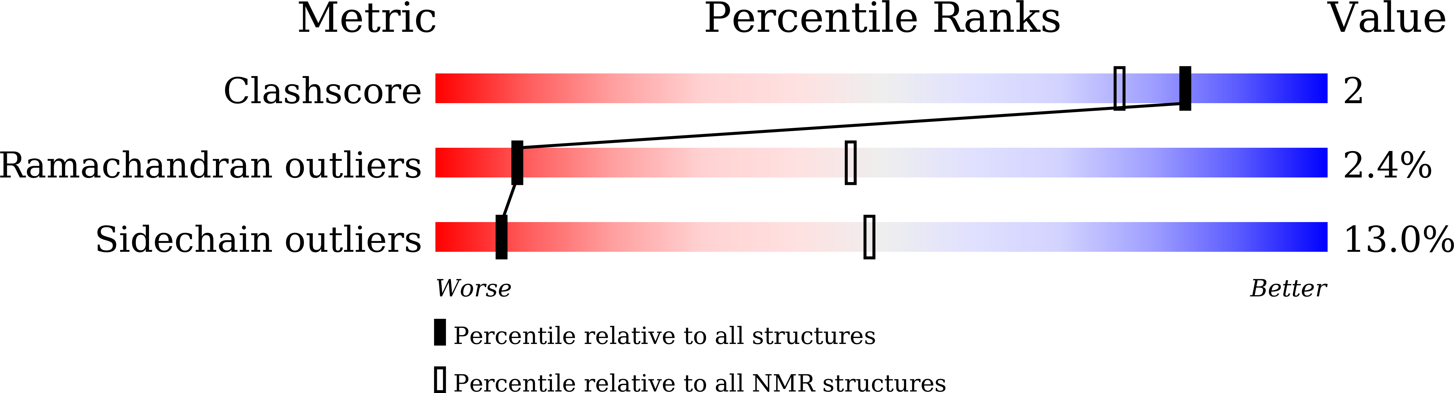
Solution structure of the mouse HOIL1-L NZF domain in the free form
Biological Source:
Source Organism:
Mus musculus (Taxon ID: 10090)
Method Details:
Experimental Method:
Conformers Calculated:
250
Conformers Submitted:
10
Selection Criteria:
structures with the least restraint violations
Macromolecular Entities
Polymer Type:polypeptide(L)
Molecule:RanBP-type and C3HC4-type zinc finger-containing protein 1
Gene (Uniprot):Rbck1
Chain IDs:A
Chain Length:66
Number of Molecules:1
Biological Source:Mus musculus
Ligand Molecules
Primary Citation
Solution structure of the HOIL-1L NZF domain reveals a conformational switch regulating linear ubiquitin affinity.
J.Biol.Chem. 299 105165 105165 (2023)
PMID: 37595872 DOI: 10.1016/j.jbc.2023.105165

Abstact

Attachment of polyubiquitin (poly-Ub) chains to proteins is a major posttranslational modification in eukaryotes. Linear ubiquitin chain assembly complex, consisting of HOIP (HOIL-1-interacting protein), HOIL-1L (heme-oxidized IRP2 Ub ligase 1), and SHARPIN (Shank-associated RH domain-interacting protein), specifically synthesizes "head-to-tail" poly-Ub chains, which are linked via the N-terminal methionine α-amino and C-terminal carboxylate of adjacent Ub units and are thus commonly called "linear" poly-Ub chains. Linear ubiquitin chain assembly complex-assembled linear poly-Ub chains play key roles in immune signaling and suppression of cell death and have been associated with immune diseases and cancer; HOIL-1L is one of the proteins known to selectively bind linear poly-Ub via its Npl4 zinc finger (NZF) domain. Although the structure of the bound form of the HOIL-1L NZF domain with linear di-Ub is known, several aspects of the recognition specificity remain unexplained. Here, we show using NMR and orthogonal biophysical methods, how the NZF domain evolves from a free to the specific linear di-Ub-bound state while rejecting other potential Ub species after weak initial binding. The solution structure of the free NZF domain revealed changes in conformational stability upon linear Ub binding, and interactions between the NZF core and tail revealed conserved electrostatic contacts, which were sensitive to charge modulation at a reported phosphorylation site: threonine-207. Phosphomimetic mutations reduced linear Ub affinity by weakening the integrity of the linear di-Ub-bound conformation. The described molecular determinants of linear di-Ub binding provide insight into the dynamic aspects of the Ub code and the NZF domain's role in full-length HOIL-1L.

Legend

Protein

Chemical

Disease

Primary Citation of related structures