8IIO image
Entry Detail
PDB ID:
8IIO
Title:
H109Q mutant of uracil DNA glycosylase X
Biological Source:
PDB Version:
Deposition Date:
2023-02-24
Release Date:
2023-06-21
Method Details:
Experimental Method:
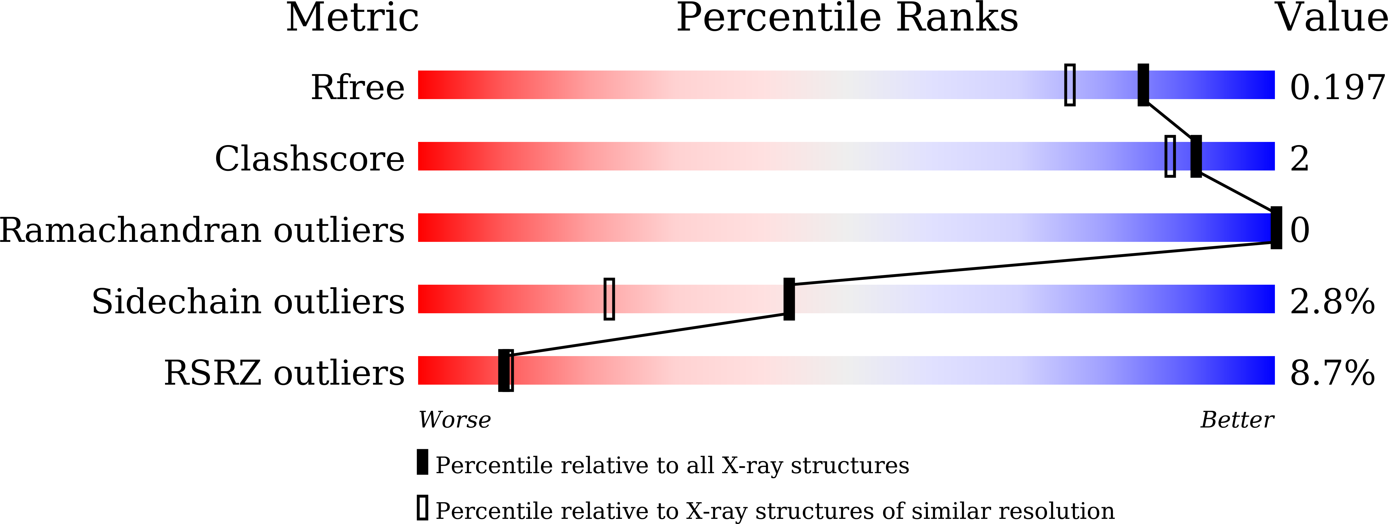
Resolution:
1.67 Å
R-Value Free:
0.19
R-Value Work:
0.18
R-Value Observed:
0.18
Space Group:
P 1 21 1
Macromolecular Entities
Polymer Type:polypeptide(L)
Description:Type-4 uracil-DNA glycosylase
Mutations:H109K
Chain IDs:A
Chain Length:207
Number of Molecules:1
Biological Source:Mycolicibacterium smegmatis MC2 155
Primary Citation
Mutational and structural analyses of UdgX: insights into the active site pocket architecture and its evolution.
Nucleic Acids Res. 51 6554 6565 (2023)
PMID: 37283083 DOI: 10.1093/nar/gkad486

Abstact

UdgX excises uracil from uracil-containing DNA to concurrently form a covalent bond with the resulting AP-DNA. Structurally, UdgX is highly similar to family-4 UDGs (F4-UDGs). However, UdgX is unique in possessing a flexible R-loop (105KRRIH109). Among the class-defining motifs, while its motif A (51GEQPG55) diverged to possess Q53 in place of A53/G53 in F4-UDGs, motif B [178HPS(S/A)(L/V)(L/V)R184] has remained unchanged. Previously, we proposed an SN1 mechanism resulting in a covalent bond between H109 and AP-DNA. In this study, we investigated several single/double mutants of UdgX. The H109A, H109S, H109G, H109Q, H109C and H109K mutants gain conventional UDG activity to varying levels. The crystal structures of UdgX mutants show topological changes in their active sites, rationalizing their UDG activities. The E52Q, E52N and E52A mutants reveal that E52 forms a catalytic dyad with H109 to enhance its nucleophilicity. The Q53A mutant supports that UdgX specific evolution of Q53 occurred essentially to stabilize the R-loop conformation. The R184A mutation (motif B) supports the role of R184 in substrate-binding. Taken together, the structural, bioinformatics, and mutational studies suggest that UdgX diverged from F4-UDGs, and the emergence of the characteristic R-loop in UdgX is functionally assisted by A53/G53 to Q53 changes in motif A.

Legend

Protein

Chemical

Disease

Primary Citation of related structures