Deposition Date
2023-02-14
Release Date
2023-05-17
Last Version Date
2024-10-16
Entry Detail
PDB ID:
8IDQ
Keywords:
Title:
Crystal structure of reducing-end xylose-releasing exoxylanase in GH30 from Talaromyces cellulolyticus with xylose
Biological Source:
Source Organism(s):
Talaromyces pinophilus (Taxon ID: 128442)
Expression System(s):
Method Details:
Experimental Method:
Resolution:
1.70 Å
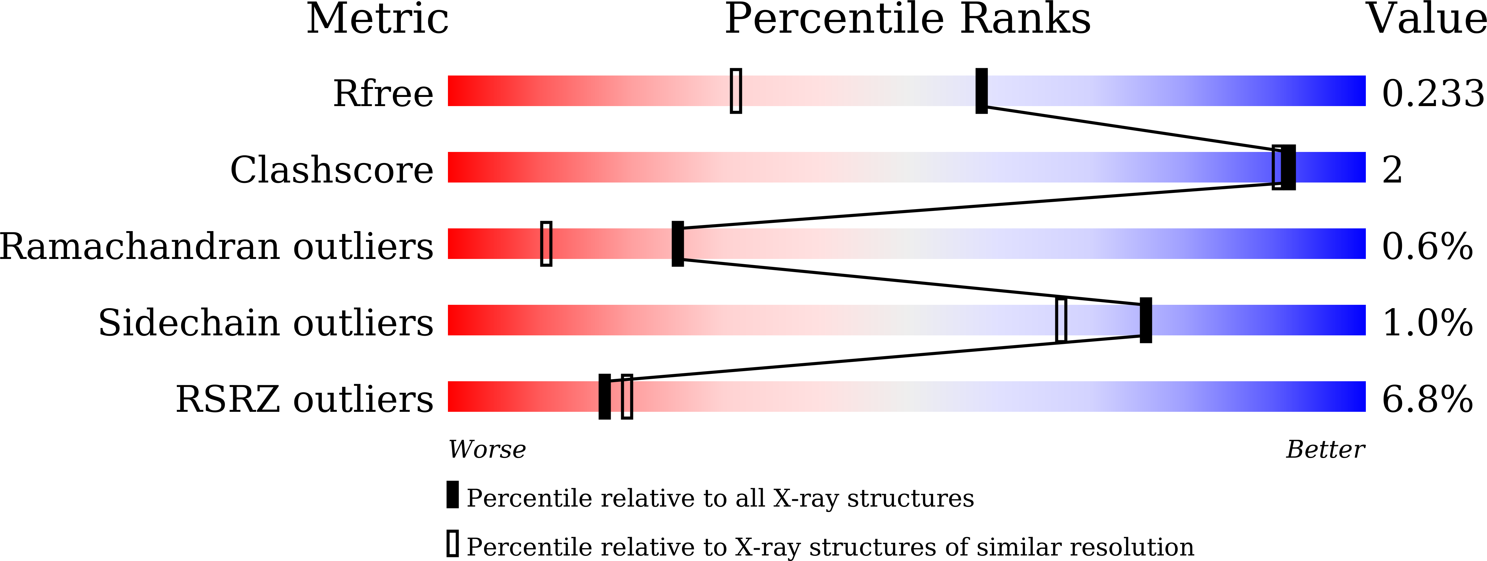
R-Value Free:
0.23
R-Value Work:
0.19
R-Value Observed:
0.20
Space Group:
P 21 21 2