
Deposition Date
2023-02-10
Release Date
2023-05-03
Last Version Date
2024-05-29
Entry Detail
PDB ID:
8IBK
Keywords:
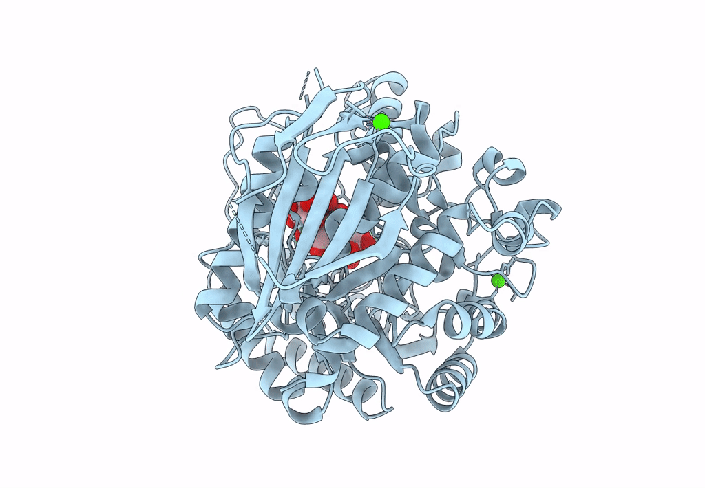
Title:
Crystal structure of Bacillus sp. AHU2216 GH13_31 Alpha-glucosidase E256Q/N258G in complex with maltotriose
Biological Source:
Source Organism(s):
Bacillus sp. (in: firmicutes) (Taxon ID: 1409)
Expression System(s):
Method Details:
Experimental Method:
Resolution:
1.69 Å
R-Value Free:
0.20
R-Value Work:
0.18
R-Value Observed:
0.18
Space Group:
P 21 21 21
