8IB0 image
Entry Detail
PDB ID:
8IB0
Keywords:
Title:
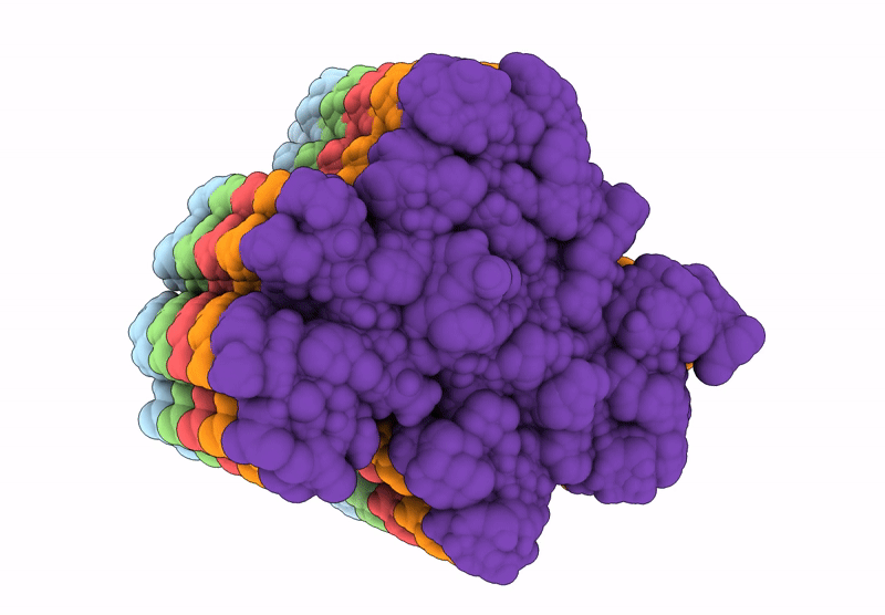
The amyloid structure of mouse RIPK1 RHIM-containing domain by solid-state NMR
Biological Source:
Source Organism:
Host Organism:
PDB Version:
Deposition Date:
2023-02-09
Release Date:
2023-03-22
Method Details:
Experimental Method:
Conformers Calculated:
196
Conformers Submitted:
10
Selection Criteria:
structures with the lowest energy
Macromolecular Entities
Polymer Type:polypeptide(L)
Description:Receptor-interacting serine/threonine-protein kinase 1
Chain IDs:A, B, C, D, E
Chain Length:24
Number of Molecules:5
Biological Source:Mus musculus
Ligand Molecules
Primary Citation
The structure of mouse RIPK1 RHIM-containing domain as a homo-amyloid and in RIPK1/RIPK3 complex.
Nat Commun 15 6975 6975 (2024)
PMID: 39143113 DOI: 10.1038/s41467-024-51303-y

Abstact

Receptor-interacting protein kinase 1 (RIPK1) is a therapeutic target in treating neurodegenerative diseases and cancers. RIPK1 has three distinct functional domains, with the center domain containing a receptor-interacting protein homotypic interaction motif (RHIM), which mediates amyloid formation. The functional amyloid formed by RIPK1 and/or RIPK3 is a crucial intermediate in regulating cell necroptosis. In this study, the amyloid structure of mouse RIPK1, formed by an 82-residue sequence centered at RHIM, is presented. It reveals the "N"-shaped folding of the protein subunit in the fibril with four β-strands. The folding pattern is shared by several amyloid structures formed by proteins with RHIM, with the central β-strand formed by the most conserved tetrad sequence I/VQI/VG. However, the solid-state NMR results indicate a structural difference between mouse RIPK1 and mouse RIPK3. A change in the structural rigidity is also suggested by the observation of weakened signals for mouse RIPK3 upon mixing with RIPK1 to form the RIPK1/RIPK3 complex fibrils. Our results provide vital information to understand the interactions between different proteins with RHIM, which will help us further comprehend the regulation mechanism in cell necroptosis.

Legend

Protein

Chemical

Disease

Primary Citation of related structures