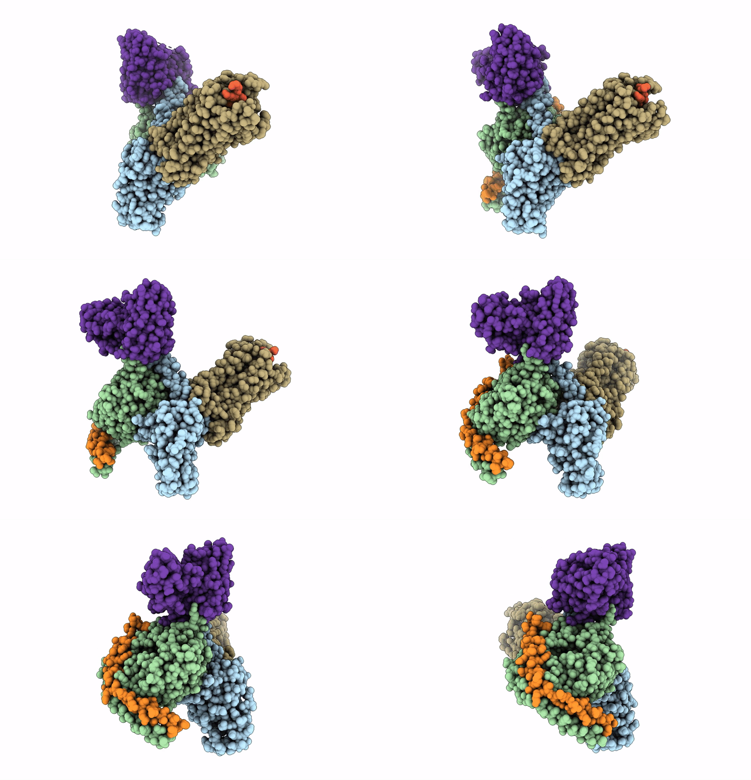
8I95 image
Deposition Date 2023-02-06
Release Date 2023-10-18
Last Version Date 2024-11-20
Entry Detail
PDB ID:
8I95
Title:
Structure of EP54-C3aR-Go complex
Biological Source:
Source Organism:
Homo sapiens (Taxon ID: 9606)
Mus musculus (Taxon ID: 10090)
Host Organism:
Method Details:
Experimental Method:
Resolution:
2.88 Å
Aggregation State:
PARTICLE
Reconstruction Method:
SINGLE PARTICLE
Macromolecular Entities
Polymer Type:polypeptide(L)
Molecule:Guanine nucleotide-binding protein G(o) subunit alpha
Gene (Uniprot):GNAO1
Mutations:G42D,E43N,A227D,G230D,I332A,V35I
Chain IDs:A
Chain Length:250
Number of Molecules:1
Biological Source:Homo sapiens
Polymer Type:polypeptide(L)
Molecule:Guanine nucleotide-binding protein G(I)/G(S)/G(T) subunit beta-1
Gene (Uniprot):GNB1
Chain IDs:B
Chain Length:350
Number of Molecules:1
Biological Source:Homo sapiens
Polymer Type:polypeptide(L)
Molecule:C3a anaphylatoxin chemotactic receptor
Gene (Uniprot):C3AR1
Chain IDs:E (auth: C)
Chain Length:538
Number of Molecules:1
Biological Source:Homo sapiens
Polymer Type:polypeptide(L)
Molecule:EP54 ligand
Chain IDs:F (auth: D)
Chain Length:10
Number of Molecules:1
Biological Source:Homo sapiens
Polymer Type:polypeptide(L)
Molecule:Guanine nucleotide-binding protein G(I)/G(S)/G(O) subunit gamma-2
Gene (Uniprot):GNG2
Chain IDs:C (auth: G)
Chain Length:71
Number of Molecules:1
Biological Source:Homo sapiens
Polymer Type:polypeptide(L)
Molecule:Antibody fragment - ScFv16
Chain IDs:D (auth: H)
Chain Length:248
Number of Molecules:1
Biological Source:Mus musculus
Primary Citation
Molecular basis of anaphylatoxin binding, activation, and signaling bias at complement receptors.
Cell 186 4956 4973.e21 (2023)
PMID: 37852260 DOI: 10.1016/j.cell.2023.09.020

Abstact

The complement system is a critical part of our innate immune response, and the terminal products of this cascade, anaphylatoxins C3a and C5a, exert their physiological and pathophysiological responses primarily via two GPCRs, C3aR and C5aR1. However, the molecular mechanism of ligand recognition, activation, and signaling bias of these receptors remains mostly elusive. Here, we present nine cryo-EM structures of C3aR and C5aR1 activated by their natural and synthetic agonists, which reveal distinct binding pocket topologies of complement anaphylatoxins and provide key insights into receptor activation and transducer coupling. We also uncover the structural basis of a naturally occurring mechanism to dampen the inflammatory response of C5a via proteolytic cleavage of the terminal arginine and the G-protein signaling bias elicited by a peptide agonist of C3aR identified here. In summary, our study elucidates the innerworkings of the complement anaphylatoxin receptors and should facilitate structure-guided drug discovery to target these receptors in a spectrum of disorders.

Legend

Protein

Chemical

Disease

Primary Citation of related structures