8I34 image
Deposition Date 2023-01-16
Release Date 2023-08-16
Last Version Date 2024-11-13
Entry Detail
PDB ID:
8I34
Title:
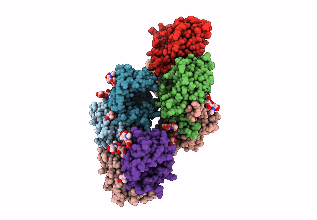
The crystal structure of EPD-BCP1 from a marine sponge
Biological Source:
Source Organism:
Haliclona sp. (Taxon ID: 34490)
Method Details:
Experimental Method:
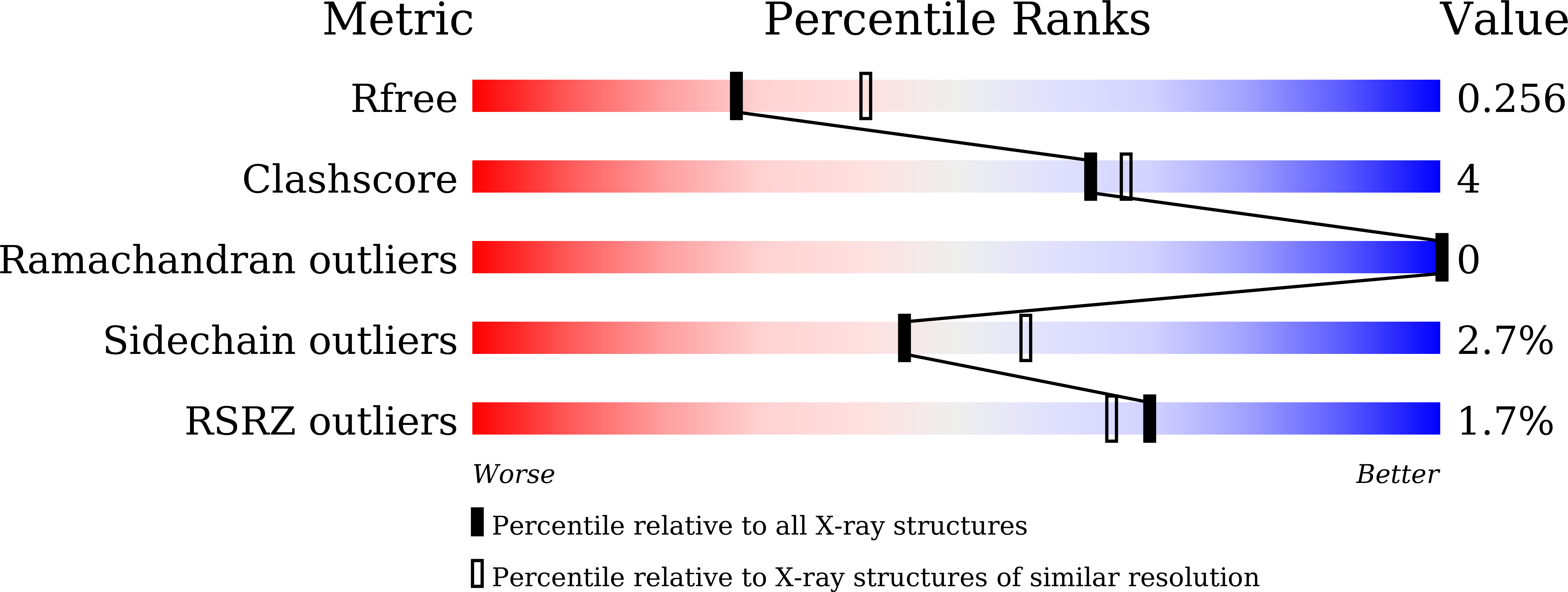
Resolution:
2.44 Å
R-Value Free:
0.24
R-Value Work:
0.20
Space Group:
P 1 21 1
Macromolecular Entities
Polymer Type:polypeptide(L)
Molecule:alpha subunit of EPD-BCP1
Chain IDs:A, C, E, G
Chain Length:196
Number of Molecules:4
Biological Source:Haliclona sp.
Polymer Type:polypeptide(L)
Molecule:beta subunit of EPD-BCP1
Chain IDs:B, D, F, H
Chain Length:196
Number of Molecules:4
Biological Source:Haliclona sp.
Primary Citation
An ependymin-related blue carotenoprotein decorates marine blue sponge.
J.Biol.Chem. 299 105110 105110 (2023)
PMID: 37517696 DOI: 10.1016/j.jbc.2023.105110

Abstact

Marine animals display diverse vibrant colors, but the mechanisms underlying their specific coloration remain to be clarified. Blue coloration is known to be achieved through a bathochromic shift of the orange carotenoid astaxanthin (AXT) by the crustacean protein crustacyanin, but other examples have not yet been well investigated. Here, we identified an ependymin (EPD)-related water-soluble blue carotenoprotein responsible for the specific coloration of the marine blue sponge Haliclona sp. EPD was originally identified in the fish brain as a protein involved in memory consolidation and neuronal regeneration. The purified blue protein, designated as EPD-related blue carotenoprotein-1, was identified as a secreted glycoprotein. We show that it consists of a heterodimer that binds orange AXT and mytiloxanthin and exhibits a bathochromic shift. Our crystal structure analysis of the natively purified EPD-related blue carotenoprotein-1 revealed that these two carotenoids are specifically bound to the heterodimer interface, where the polyene chains are aligned in parallel to each other like in β-crustacyanin, although the two proteins are evolutionary and structurally unrelated. Furthermore, using reconstitution assays, we found that incomplete bathochromic shifts occurred when the protein bound to only AXT or mytiloxanthin. Taken together, we identified an EPD in a basal metazoan as a blue protein that decorates the sponge body by binding specific structurally unrelated carotenoids.

Legend

Protein

Chemical

Disease

Primary Citation of related structures