Deposition Date
2023-01-13
Release Date
2023-03-22
Last Version Date
2024-05-29
Entry Detail
PDB ID:
8I1D
Keywords:
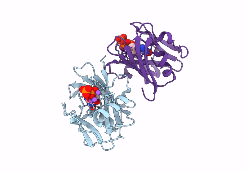
Title:
Crystal structure of human MTH1(G2K mutant) in complex with 2-oxo-dATP at pH 7.7
Biological Source:
Source Organism(s):
Homo sapiens (Taxon ID: 9606)
Expression System(s):
Method Details:
Experimental Method:
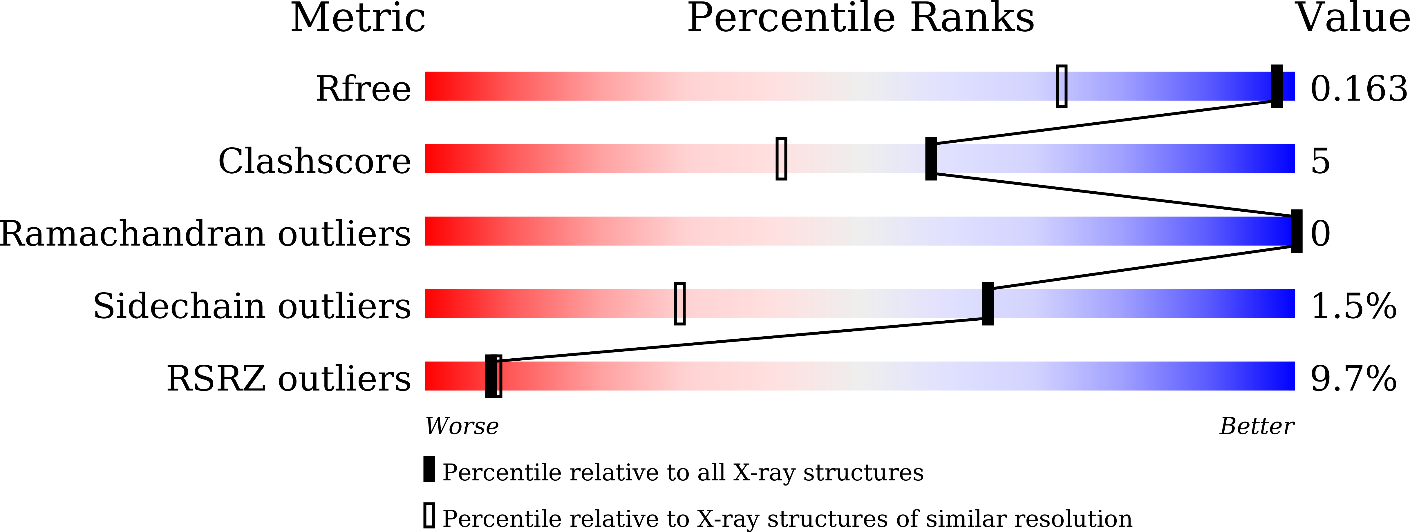
Resolution:
1.20 Å
R-Value Free:
0.16
R-Value Work:
0.13
R-Value Observed:
0.13
Space Group:
P 21 21 21