8HVB image
Deposition Date 2022-12-26
Release Date 2023-12-27
Last Version Date 2025-07-09
Entry Detail
PDB ID:
8HVB
Keywords:
Title:
Crystal structure of lacto-N-biosidase StrLNBase from Streptomyces sp. strain 142, lacto-N-biose complex
Biological Source:
Source Organism:
Method Details:
Experimental Method:
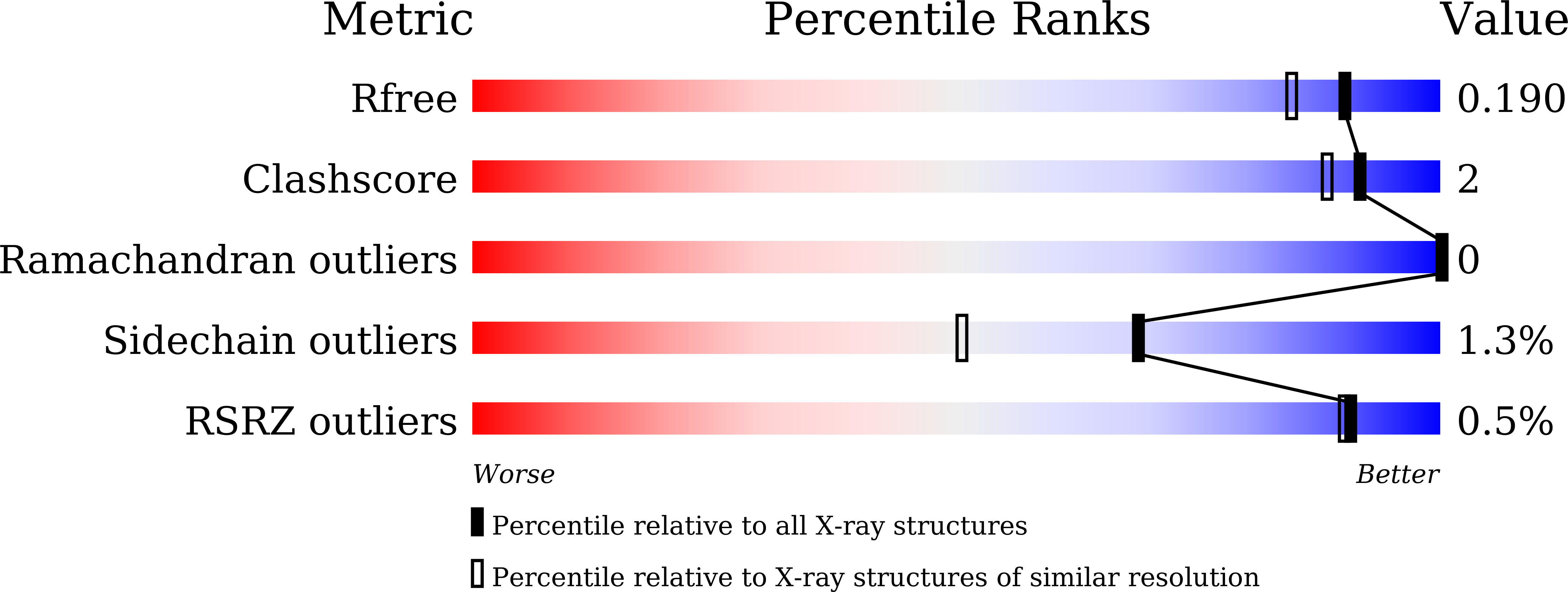
Resolution:
1.60 Å
R-Value Free:
0.18
R-Value Work:
0.14
R-Value Observed:
0.15
Space Group:
P 1 21 1
Macromolecular Entities
Polymer Type:polypeptide(L)
Molecule:Lacto-N-biosidase
Chain IDs:A
Chain Length:614
Number of Molecules:1
Biological Source:Streptomyces sp.
Primary Citation
Crystal Structure of Glycoside Hydrolase Family 20 Lacto- N -biosidase from Soil Bacterium Streptomyces sp. Strain 142.
J Appl Glycosci (1999) 72 7202101 7202101 (2025)
PMID: 40502377 DOI: 10.5458/jag.7202101

Abstact

Lacto-N-biosidase hydrolyzes the β-GlcNAc or β-GalNAc bond of sugar chains to release lacto-N-biose I (Gal-β1,3-GlcNAc) or galacto-N-biose (Gal-β1,3-GalNAc) from the non-reducing end. Typical substrates for lacto-N-biosidase include type I oligosaccharides contained in human breast milk, such as lacto-N-tetraose. Lacto-N-biosidases have recently received significant attention because of their potential to synthesize milk oligosaccharides. Bifidobacterial lacto-N-biosidases belonging to glycoside hydrolase families 20 and 136 have been studied. The GH20 lacto-N-biosidases utilize a substrate-associated hydrolysis mechanism. LnbB from Bifidobacterium bifidum is the only lacto-N-biosidase with reported crystal structures in GH20. In this study, the crystal structure of the lacto-N-biosidase from Streptomyces sp. strain 142 (StrLNBase) was solved in a complex with lacto-N-biose and galacto-N-biose. The stabilizing residue, which recognizes the nitrogen atom of the N-acetyl group of the -1 subsite, and the catalytic acid/base residue, were determined to be D304 and E305, respectively. The structure of StrLNBase is similar to that of LnbB; however, in the complex with galacto-N-biose, there were two structures exhibiting different sugar conformations. A phylogenetic analysis revealed that lacto-N-biosidases discovered in the soil bacteria Streptomyces spp. and human gut bacteria Bifidobacterium spp. may be divided into two separate groups, which suggests that they evolved divergently.

Legend

Protein

Chemical

Disease

Primary Citation of related structures