8HU7 image
Entry Detail
PDB ID:
8HU7
Title:
Crystal structure of FGF2-M1 mutant - D28E/C78L/C96I/S137P
Biological Source:
Source Organism:
Host Organism:
PDB Version:
Deposition Date:
2022-12-22
Release Date:
2024-06-26
Method Details:
Experimental Method:
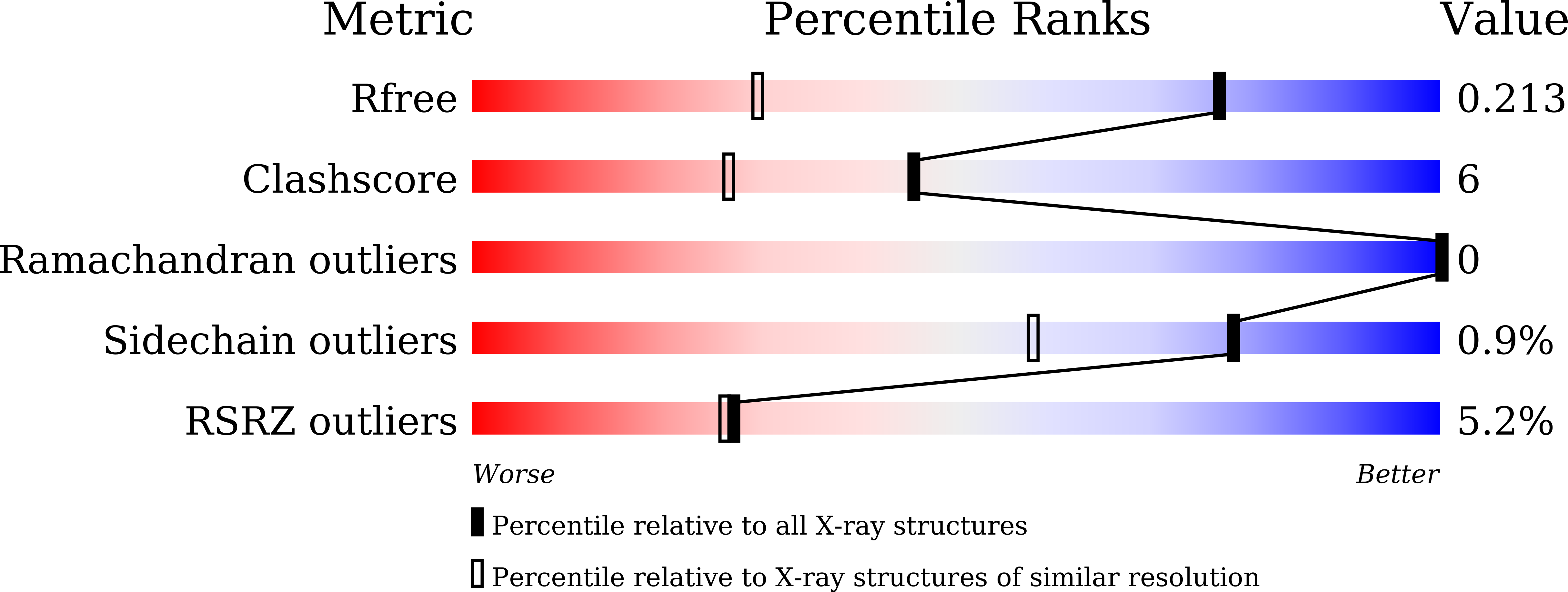
Resolution:
1.40 Å
R-Value Free:
0.21
R-Value Work:
0.18
Space Group:
P 31 2 1
Macromolecular Entities
Polymer Type:polypeptide(L)
Description:Fibroblast growth factor 2
Mutations:D28E/C78I/C96I/S137P
Chain IDs:A
Chain Length:147
Number of Molecules:1
Biological Source:Homo sapiens
Primary Citation
Structural and biochemical investigation into stable FGF2 mutants with novel mutation sites and hydrophobic replacements for surface-exposed cysteines.
Plos One 19 e0307499 e0307499 (2024)
PMID: 39236042 DOI: 10.1371/journal.pone.0307499

Abstact

Fibroblast growth factor 2 (FGF2) is an attractive biomaterial for pharmaceuticals and functional cosmetics. To improve the thermo-stability of FGF2, we designed two mutants harboring four-point mutations: FGF2-M1 (D28E/C78L/C96I/S137P) and FGF2-M2 (D28E/C78I/C96I/S137P) through bioinformatics, molecular thermodynamics, and molecular modeling. The D28E mutation reduced fragmentation of the FGF2 wild type during preparation, and the substitution of a whale-specific amino acid, S137P, enhanced the thermal stability of FGF2. Surface-exposed cysteines that participate in oligomerization through intermolecular disulfide bond formation were substituted with hydrophobic residues (C78L/C78I and C96I) using the in silico method. High-resolution crystal structures revealed at the atomic level that the introduction of mutations stabilizes each local region by forming more favorable interactions with neighboring residues. In particular, P137 forms CH-π interactions with the side chain indole ring of W123, which seems to stabilize a β-hairpin structure, containing a heparin-binding site of FGF2. Compared to the wild type, both FGF2-M1 and FGF2-M2 maintained greater solubility after a week at 45 °C, with their Tm values rising by ~ 5 °C. Furthermore, the duration for FGF2-M1 and FGF2-M2 to reach 50% residual activity at 45 °C extended to 8.8- and 8.2-fold longer, respectively, than that of the wild type. Interestingly, the hydrophobic substitution of surface-exposed cysteine in both FGF2 mutants makes them more resistant to proteolytic cleavage by trypsin, subtilisin, proteinase K, and actinase than the wild type and the Cys → Ser substitution. The hydrophobic replacements can influence protease resistance as well as oligomerization and thermal stability. It is notable that hydrophobic substitutions of surface-exposed cysteines, as well as D28E and S137P of the FGF2 mutants, were designed through various approaches with structural implications. Therefore, the engineering strategies and structural insights adopted in this study could be applied to improve the stability of other proteins.

Legend

Protein

Chemical

Disease

Primary Citation of related structures