Deposition Date
2022-12-20
Release Date
2023-07-19
Last Version Date
2024-05-29
Entry Detail
PDB ID:
8HSV
Keywords:
Title:
The structure of rat beta-arrestin1 in complex with a rat Mdm2 peptide
Biological Source:
Source Organism(s):
Rattus norvegicus (Taxon ID: 10116)
Expression System(s):
Method Details:
Experimental Method:
Resolution:
3.00 Å
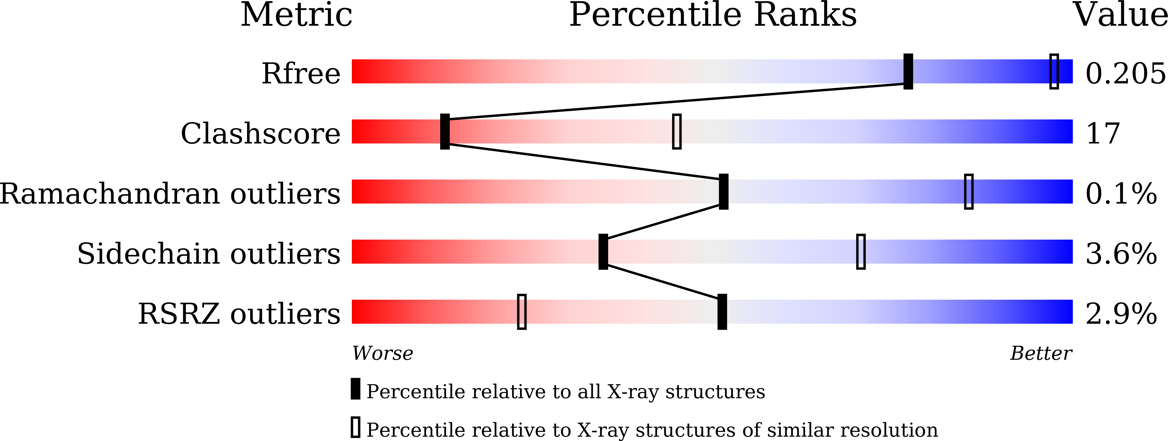
R-Value Free:
0.20
R-Value Work:
0.17
R-Value Observed:
0.17
Space Group:
P 32