8HOV image
Deposition Date 2022-12-11
Release Date 2024-06-12
Last Version Date 2025-01-15
Entry Detail
PDB ID:
8HOV
Title:
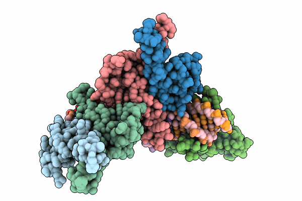
Crystal structure of Hms1p from Saccharomyces cerevisiae
Biological Source:
Source Organism:
Host Organism:
Method Details:
Experimental Method:
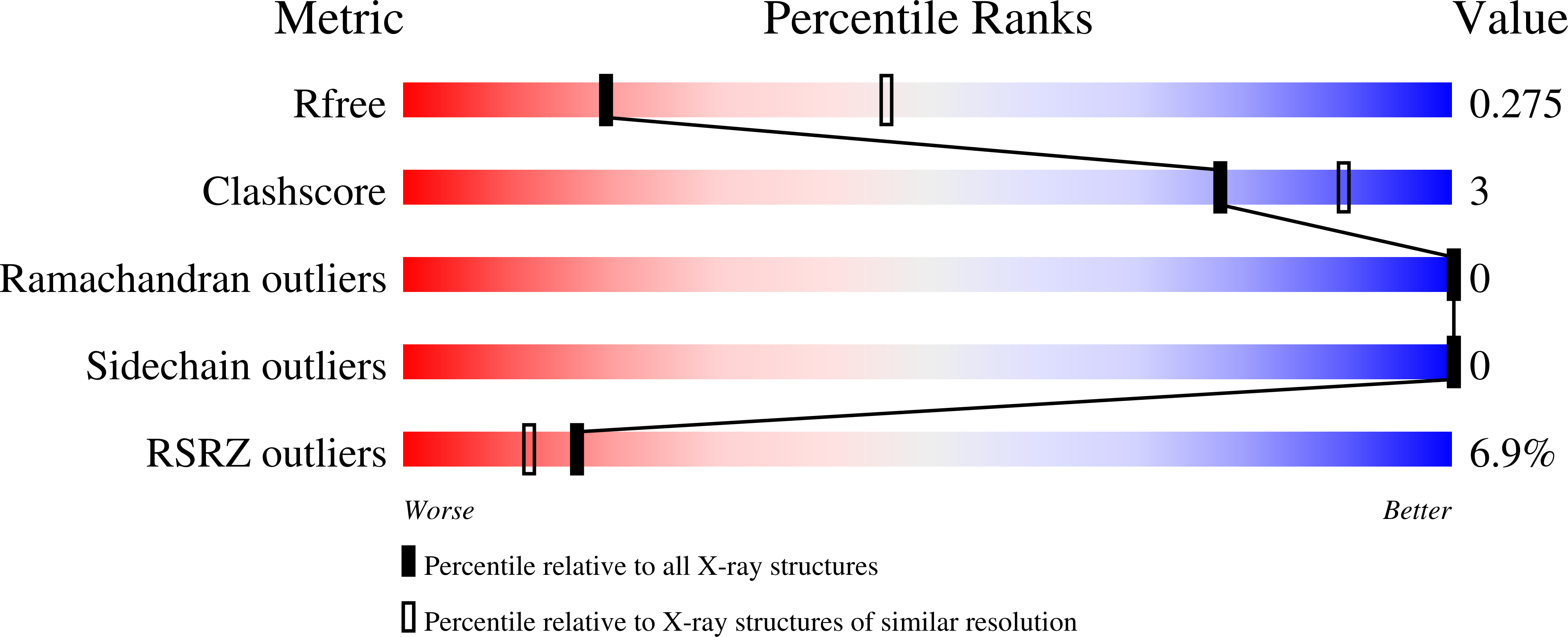
Resolution:
2.77 Å
R-Value Free:
0.27
R-Value Work:
0.23
R-Value Observed:
0.23
Space Group:
P 1 21 1
Macromolecular Entities
Structures with similar UniProt ID
Protein Blast
Polymer Type:polypeptide(L)
Molecule:Transcription factor HMS1
Gene (Uniprot):HMS1
Chain IDs:A, B, C, D, E, F
Chain Length:108
Number of Molecules:6
Biological Source:Saccharomyces cerevisiae
Polymer Type:polydeoxyribonucleotide
Molecule:DNA (5'-D(*TP*CP*AP*CP*GP*CP*AP*T)-3')
Chain IDs:G, H, K
Chain Length:8
Number of Molecules:3
Biological Source:Saccharomyces cerevisiae
Polymer Type:polydeoxyribonucleotide
Molecule:DNA (5'-D(*AP*TP*GP*CP*GP*TP*GP*A)-3')
Chain IDs:I, J, L
Chain Length:8
Number of Molecules:3
Biological Source:Saccharomyces cerevisiae
Primary Citation
Molecular recognition of the promoter DNA signature sequence by Hms1p DBD.
Int.J.Biol.Macromol. ? 139232 139232 (2025)
PMID: 39756762 DOI: 10.1016/j.ijbiomac.2024.139232

Abstact

Transcriptional regulation of sterol biosynthetic genes is mediated by conserved sterol-regulatory element binding proteins (SREBPs) in human pathogenic fungi, however, its homolog in S. cerevisiae regulate filamentous growth during stress conditions. These pseudohyphal growths might be associated with the expression of MEP2 gene in response to ammonium limitation. Hitherto, there is limited literature available for Hms1p and precisely how it establishes interaction with DNA. Though DNA and Hms1p mutual interaction was predicted computationally, however, the structural details regarding how they establish interaction still remains elusive. Here, we resolved the crystal structure of Hms1pDBD-DNA complex at a nominal resolution of 2.77 Å. The structure highlighted several residues (Hms1pHis3/Asn4/Glu7/Tyr10/Arg11) could specifically recognize the core signature sequence in the promoter DNA fragment, which was validated by biochemical assays. Comparative analysis of Hms1p with other basic helix-loop-helix (bHLH) transcriptional regulators reflected that residues (His, Glu and Arg) are highly conserved. Despite distinct core signature sequences, these conserved residues in different bHLH proteins could specifically recognize and bind their corresponding promoter DNA fragment. Collectively, these results could pinpoint critical residues (Hms1pHis3/Asn4/Glu7/Tyr10/Arg11) for the binding interface with the signature sequence of MEP2 promoter DNA fragment.

Legend

Protein

Chemical

Disease

Primary Citation of related structures
Feedback Form
Name
Email
Institute
Feedback