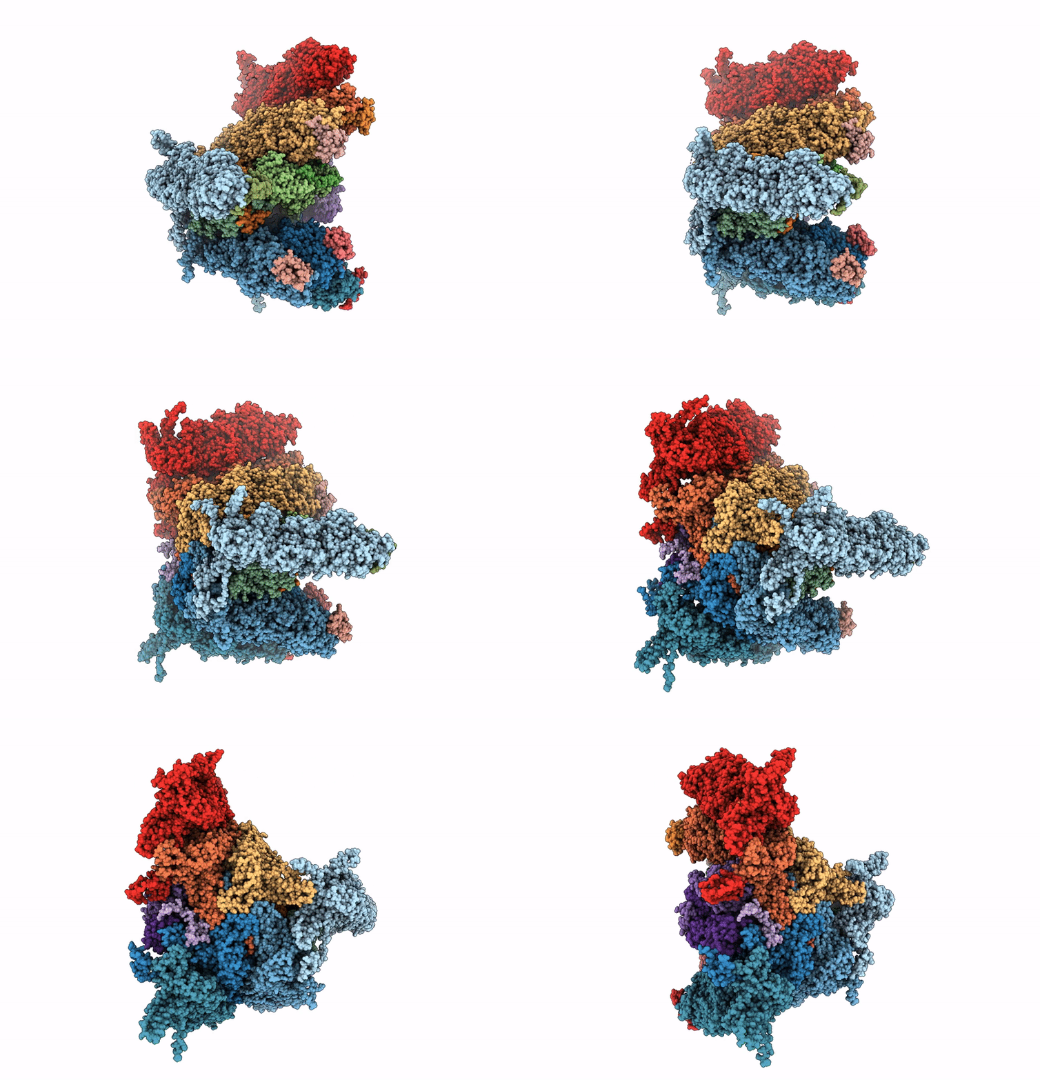
8HEY image
Deposition Date 2022-11-09
Release Date 2023-11-01
Last Version Date 2025-06-25
Entry Detail
PDB ID:
8HEY
Keywords:
Title:
One CVSC-binding penton vertex in HCMV B-capsid
Biological Source:
Source Organism:
Method Details:
Experimental Method:
Resolution:
4.10 Å
Aggregation State:
PARTICLE
Reconstruction Method:
SINGLE PARTICLE
Macromolecular Entities
Polymer Type:polypeptide(L)
Molecule:Capsid vertex component 1
Chain IDs:T (auth: M)
Chain Length:594
Number of Molecules:1
Biological Source:Human betaherpesvirus 5
Polymer Type:polypeptide(L)
Molecule:Capsid vertex component 2
Chain IDs:U (auth: N), V (auth: O)
Chain Length:306
Number of Molecules:2
Biological Source:Human betaherpesvirus 5
Polymer Type:polypeptide(L)
Molecule:Small capsomere-interacting protein
Gene (Uniprot):UL48A
Chain IDs:A (auth: T), B (auth: i), C (auth: j), N (auth: Q), O (auth: R), P (auth: S)
Chain Length:306
Number of Molecules:6
Biological Source:Human betaherpesvirus 5
Polymer Type:polypeptide(L)
Molecule:Major capsid protein
Gene (Uniprot):UL86
Chain IDs:D (auth: a), E (auth: D), F (auth: Y), G (auth: Z), Q (auth: A), R (auth: B), S (auth: C)
Chain Length:1370
Number of Molecules:7
Biological Source:Human betaherpesvirus 5
Polymer Type:polypeptide(L)
Molecule:Triplex capsid protein 1
Gene (Uniprot):UL46
Chain IDs:L (auth: g), M (auth: m)
Chain Length:290
Number of Molecules:2
Biological Source:Human betaherpesvirus 5
Polymer Type:polypeptide(L)
Molecule:Triplex capsid protein 2
Gene (Uniprot):UL85
Chain IDs:H (auth: h), I, J (auth: n), K (auth: o)
Chain Length:306
Number of Molecules:4
Biological Source:Human betaherpesvirus 5
Ligand Molecules
Primary Citation
Cryo-electron microscopy structures of capsids and in situ portals of DNA-devoid capsids of human cytomegalovirus.
Nat Commun 14 2025 2025 (2023)
PMID: 37041152 DOI: 10.1038/s41467-023-37779-0

Abstact

The portal-scaffold complex is believed to nucleate the assembly of herpesvirus procapsids. During capsid maturation, two events occur: scaffold expulsion and DNA incorporation. The portal-scaffold interaction and the conformational changes that occur to the portal during the different stages of capsid formation have yet to be elucidated structurally. Here we present high-resolution structures of the A- and B-capsids and in-situ portals of human cytomegalovirus. We show that scaffolds bind to the hydrophobic cavities formed by the dimerization and Johnson-fold domains of the major capsid proteins. We further show that 12 loop-helix-loop fragments-presumably from the scaffold domain-insert into the hydrophobic pocket of the portal crown domain. The portal also undergoes significant changes both positionally and conformationally as it accompanies DNA packaging. These findings unravel the mechanism by which the portal interacts with the scaffold to nucleate capsid assembly and further our understanding of scaffold expulsion and DNA incorporation.

Legend

Protein

Chemical

Disease

Primary Citation of related structures