Home
About
Browse
Covid-19
Organism Classification
Alphafold Collection
Virus Classification
Indian Data
Contact
Search Type
PDB Id
Uniprot Id
Source Organism
Host Organism
Protein Name
Method
Summary
Structure Feature
Experiment
Ligands & Environment
8HEO
pdb_00008heo
10.2210/pdb8heo/pdb
Download file
FASTA
PDB
MMCIF
Binary MMCIF
XML
Structure Factors
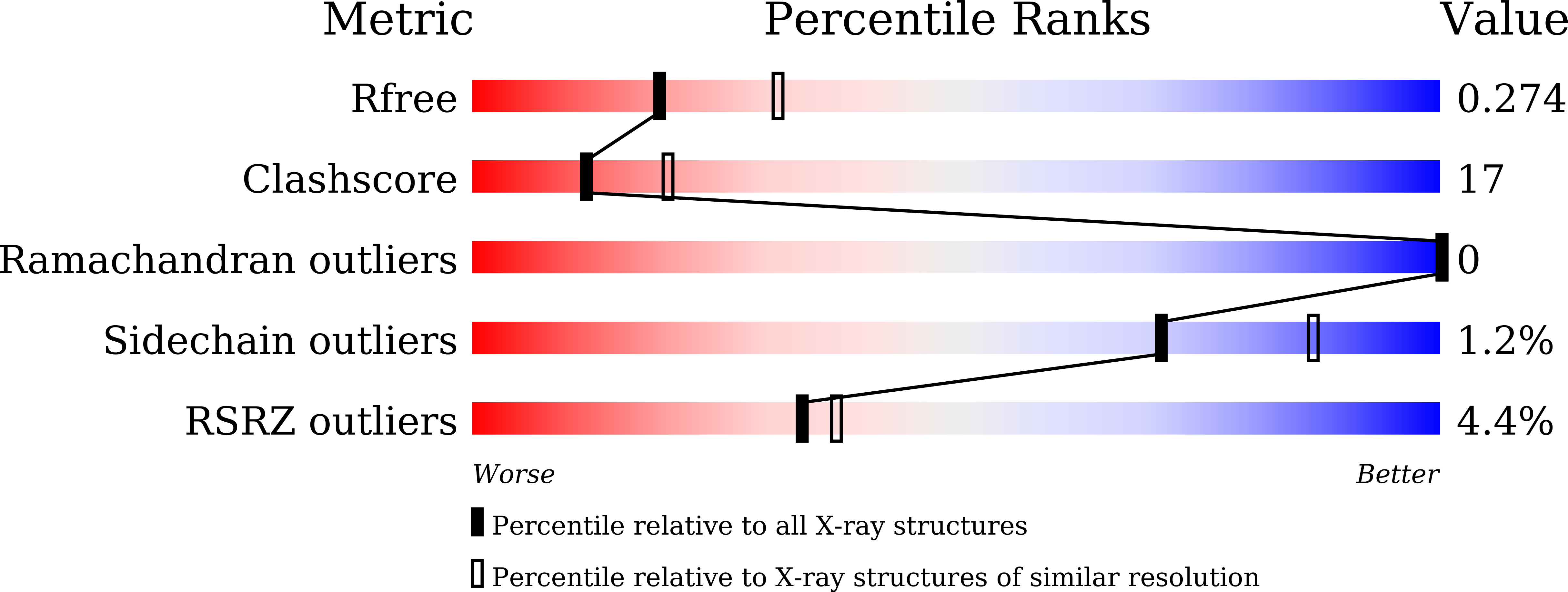
Full Validation Report
Validation File (XML)
Validation File (CIF)
FASTA Zipped(.gz)
PDB Zipped(.gz)
MMCIF Zipped(.gz)
Binary MMCIF Zipped(.gz)
Structure Factors Zipped(.gz)
Validation File Zipped (.xml.gz)
Validation File Zipped (.cif.gz)
Deposition Date
2022-11-08
Release Date
2023-11-15
Last Version Date
2023-11-15
Entry Detail
PDB ID:
8HEO
Keywords:
LIGASE
Title:
Crystal structure of SIAH1 SBD bound to Axin2 peptide
Biological Source:
Source Organism:
Homo sapiens
(Taxon ID:
9606
)
Host Organism:
Escherichia coli BL21(DE3)
Method Details:
Experimental Method:
X-RAY DIFFRACTION
Resolution:
2.53 Å
R-Value Work:
0.20
Space Group:
P 1 21 1
Download Validation Report
Macromolecular Entities
Structures with similar UniProt ID
Protein Blast
Copy Sequence
Download FASTA
Polymer Type:
polypeptide(L)
Molecule:
E3 ubiquitin-protein ligase SIAH1
Gene (Uniprot):
SIAH1
Chain IDs:
A
,
B
Chain Length:
194
Number of Molecules:
2
Biological Source:
Homo sapiens
Structures with similar UniProt ID
Protein Blast
Copy Sequence
Download FASTA
Polymer Type:
polypeptide(L)
Molecule:
Peptide from Axin-2
Gene (Uniprot):
AXIN2
Chain IDs:
C
(auth: G)
Chain Length:
21
Number of Molecules:
1
Biological Source:
Homo sapiens
Ligand Molecules
ZN
Ligand Environment View
Primary Citation
Crystal structure of SIAH1 SBD bound to Axin2 peptide
Yan, X.X
,
Tian, L.F.
Show
To Be Published ? ? ? (?)
Primary Citation of related structures
8HEO