8HDR image
Entry Detail
PDB ID:
8HDR
EMDB ID:
Keywords:
Title:
Cyanophage Pam3 neck
Biological Source:
Source Organism:
PDB Version:
Deposition Date:
2022-11-06
Release Date:
2023-02-01
Method Details:
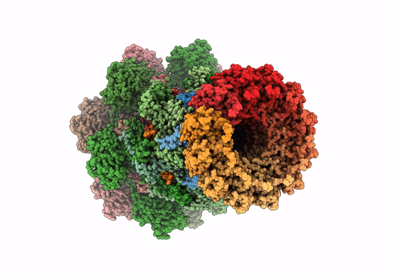
Experimental Method:
Resolution:
3.66 Å
Aggregation State:
PARTICLE
Reconstruction Method:
SINGLE PARTICLE
Macromolecular Entities
Polymer Type:polypeptide(L)
Description:pam3 tube protein
Chain IDs:A (auth: 1), B (auth: 2), AA (auth: Y), BA (auth: Z), OA (auth: m), PA (auth: n), QA (auth: o), RA (auth: p), SA (auth: q), TA (auth: r), UA (auth: s), VA (auth: t), WA (auth: u), XA (auth: v), YA (auth: w), ZA (auth: x), AB (auth: y), BB (auth: z)
Chain Length:142
Number of Molecules:18
Biological Source:uncultured cyanophage
Polymer Type:polypeptide(L)
Description:Pam3 connector protein
Chain IDs:C (auth: A), D (auth: B), E (auth: C), F (auth: D), G (auth: E), H (auth: F)
Chain Length:131
Number of Molecules:6
Biological Source:uncultured cyanophage
Polymer Type:polypeptide(L)
Description:Pam3 terminator protein
Chain IDs:I (auth: G), J (auth: H), K (auth: I), L (auth: J), M (auth: K), N (auth: L)
Chain Length:131
Number of Molecules:6
Biological Source:uncultured cyanophage
Polymer Type:polypeptide(L)
Description:Pam3 sheath protein
Chain IDs:O (auth: M), P (auth: N), Q (auth: O), R (auth: P), S (auth: Q), T (auth: R), U (auth: S), V (auth: T), W (auth: U), X (auth: V), Y (auth: W), Z (auth: X)
Chain Length:384
Number of Molecules:12
Biological Source:uncultured cyanophage
Polymer Type:polypeptide(L)
Description:Pam3 adaptor protein
Chain IDs:CA (auth: a), DA (auth: b), EA (auth: c), FA (auth: d), GA (auth: e), HA (auth: f), IA (auth: g), JA (auth: h), KA (auth: i), LA (auth: j), MA (auth: k), NA (auth: l)
Chain Length:131
Number of Molecules:12
Biological Source:uncultured cyanophage
Ligand Molecules
Primary Citation
Fine structure and assembly pattern of a minimal myophage Pam3.
Proc.Natl.Acad.Sci.USA 120 e2213727120 e2213727120 (2023)
PMID: 36656854 DOI: 10.1073/pnas.2213727120

Abstact

The myophage possesses a contractile tail that penetrates its host cell envelope. Except for investigations on the bacteriophage T4 with a rather complicated structure, the assembly pattern and tail contraction mechanism of myophage remain largely unknown. Here, we present the fine structure of a freshwater Myoviridae cyanophage Pam3, which has an icosahedral capsid of ~680 Å in diameter, connected via a three-section neck to an 840-Å-long contractile tail, ending with a three-module baseplate composed of only six protein components. This simplified baseplate consists of a central hub-spike surrounded by six wedge heterotriplexes, to which twelve tail fibers are covalently attached via disulfide bonds in alternating upward and downward configurations. In vitro reduction assays revealed a putative redox-dependent mechanism of baseplate assembly and tail sheath contraction. These findings establish a minimal myophage that might become a user-friendly chassis phage in synthetic biology.

Legend

Protein

Chemical

Disease

Primary Citation of related structures